3RO4 | pdb_00003ro4

X-ray Structure of Ketohexokinase in complex with an indazole compound derivative


Experimental Data Snapshot

  • Method: X-RAY DIFFRACTION
  • Resolution: 2.60 Å
  • R-Value Free: 
    0.297 (Depositor), 0.255 (DCC) 
  • R-Value Work: 
    0.258 (Depositor), 0.214 (DCC) 
  • R-Value Observed: 
    0.261 (Depositor) 

Starting Model: experimental
View more details

wwPDB Validation 3D Report Full Report

Validation slider image for 3RO4

Ligand Structure Quality Assessment 


This is version 1.4 of the entry. See complete history

Literature

Optimization of a pyrazole hit from FBDD into a novel series of indazoles as ketohexokinase inhibitors.

Zhang, X.Song, F.Kuo, G.H.Xiang, A.Gibbs, A.C.Abad, M.C.Sun, W.Kuo, L.C.Sui, Z.

(2011) Bioorg Med Chem Lett 21: 4762-4767

  • DOI: https://doi.org/10.1016/j.bmcl.2011.06.067
  • Primary Citation Related Structures: 
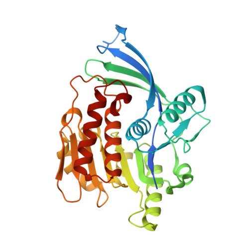
    3RO4

  • PubMed Abstract: 

    A series of indazoles have been discovered as KHK inhibitors from a pyrazole hit identified through fragment-based drug discovery (FBDD). The optimization process guided by both X-ray crystallography and solution activity resulted in lead-like compounds with good pharmaceutical properties.


  • Organizational Affiliation
    • Johnson & Johnson Pharmaceutical Research and Development, Welsh & McKean Roads, PO Box 776, Spring House, PA 19477, United States. xzhang5@its.jnj.com

Macromolecule Content 

  • Total Structure Weight: 68.68 kDa 
  • Atom Count: 4,545 
  • Modeled Residue Count: 587 
  • Deposited Residue Count: 626 
  • Unique protein chains: 1

Macromolecules

Find similar proteins by:|  3D Structure
Entity ID: 1
MoleculeChains  Sequence LengthOrganismDetailsImage
Ketohexokinase
A, B
313Homo sapiensMutation(s): 0 
Gene Names: KHK
EC: 2.7.1.3
UniProt & NIH Common Fund Data Resources
Find proteins for P50053 (Homo sapiens)
Explore P50053 
Go to UniProtKB:  P50053
PHAROS:  P50053
GTEx:  ENSG00000138030 
Entity Groups
Sequence Clusters30% Identity50% Identity70% Identity90% Identity95% Identity100% Identity
UniProt GroupP50053
Sequence Annotations
Expand
Reference Sequence

Experimental Data & Validation

Experimental Data

  • Method: X-RAY DIFFRACTION
  • Resolution: 2.60 Å
  • R-Value Free:  0.297 (Depositor), 0.255 (DCC) 
  • R-Value Work:  0.258 (Depositor), 0.214 (DCC) 
  • R-Value Observed: 0.261 (Depositor) 
Space Group: P 21 21 21
Unit Cell:
Length ( Å )Angle ( ˚ )
a = 83.61α = 90
b = 86.034β = 90
c = 139.792γ = 90
Software Package:
Software NamePurpose
JDirectordata collection
PHENIXmodel building
PHENIXrefinement
d*TREKdata reduction
d*TREKdata scaling
PHENIXphasing

Structure Validation

View Full Validation Report


Ligand Structure Quality Assessment 


Entry History 

Deposition Data

Revision History  (Full details and data files)

  • Version 1.0: 2011-08-03
    Type: Initial release
  • Version 1.1: 2012-05-23
    Changes: Database references
  • Version 1.2: 2012-12-05
    Changes: Other
  • Version 1.3: 2017-11-08
    Changes: Refinement description
  • Version 1.4: 2023-09-13
    Changes: Data collection, Database references, Derived calculations, Refinement description