3RLK | pdb_00003rlk

Crystal structure of the read-through domain from bacteriophage Qbeta A1 protein, monoclinic crystal form


Experimental Data Snapshot

  • Method: X-RAY DIFFRACTION
  • Resolution: 1.76 Å
  • R-Value Free: 
    0.242 (Depositor), 0.242 (DCC) 
  • R-Value Work: 
    0.174 (Depositor), 0.174 (DCC) 
  • R-Value Observed: 
    0.177 (Depositor) 

wwPDB Validation 3D Report Full Report

Validation slider image for 3RLK

This is version 1.2 of the entry. See complete history

Literature

Crystal structure of the read-through domain from bacteriophage Qbeta A1 protein

Rumnieks, J.Tars, K.

(2011) Protein Sci 20: 1707-1712

  • DOI: https://doi.org/10.1002/pro.704
  • Primary Citation Related Structures: 
    3RLC, 3RLK

  • PubMed Abstract: 

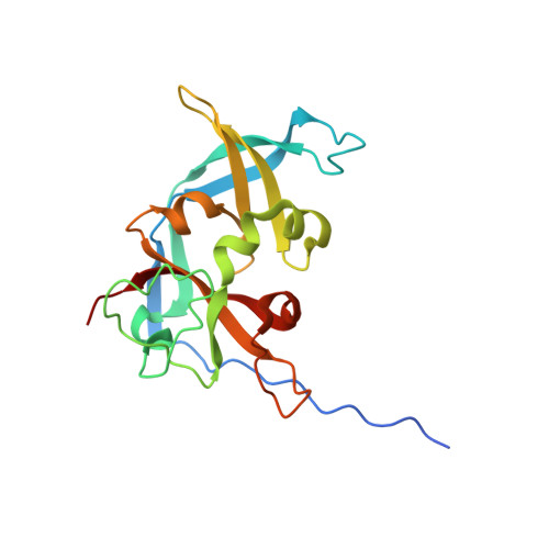
    Bacteriophage Qβ is a small RNA virus that infects Escherichia coli. The virus particle contains a few copies of the minor coat protein A1, a C-terminally prolonged version of the coat protein, which is formed when ribosomes occasionally read-through the leaky stop codon of the coat protein. The crystal structure of the read-through domain from bacteriophage Qβ A1 protein was determined at a resolution of 1.8 Å. The domain consists of a heavily deformed five-stranded β-barrel on one side of the protein and a β-hairpin and a three-stranded β-sheet on the other. Several short helices and well-ordered loops are also present throughout the protein. The N-terminal part of the read-through domain contains a prominent polyproline type II helix. The overall fold of the domain is not similar to any published structure in the Protein Data Bank.


  • Organizational Affiliation
    • Latvian Biomedical Research and Study Centre, Ratsupites 1, Riga LV-1067, Latvia. j.rumnieks@biomed.lu.lv

Macromolecule Content 

  • Total Structure Weight: 21.99 kDa 
  • Atom Count: 1,631 
  • Modeled Residue Count: 183 
  • Deposited Residue Count: 192 
  • Unique protein chains: 1

Macromolecules

Find similar proteins by:|  3D Structure
Entity ID: 1
MoleculeChains  Sequence LengthOrganismDetailsImage
A1 protein192Qubevirus durumMutation(s): 0 
Gene Names: A1
UniProt
Find proteins for Q8LTE1 (Escherichia virus Qbeta)
Explore Q8LTE1 
Go to UniProtKB:  Q8LTE1
Entity Groups
Sequence Clusters30% Identity50% Identity70% Identity90% Identity95% Identity100% Identity
UniProt GroupQ8LTE1
Sequence Annotations
Expand
Reference Sequence

Small Molecules

Ligands 1 Unique
IDChains Name / Formula / InChI Key2D Diagram3D Interactions
PG4

Query on PG4


Download:Ideal Coordinates CCD File
B [auth A]TETRAETHYLENE GLYCOL
C8 H18 O5
UWHCKJMYHZGTIT-UHFFFAOYSA-N

Experimental Data & Validation

Experimental Data

  • Method: X-RAY DIFFRACTION
  • Resolution: 1.76 Å
  • R-Value Free:  0.242 (Depositor), 0.242 (DCC) 
  • R-Value Work:  0.174 (Depositor), 0.174 (DCC) 
  • R-Value Observed: 0.177 (Depositor) 
Space Group: P 1 21 1
Unit Cell:
Length ( Å )Angle ( ˚ )
a = 44.01α = 90
b = 49.12β = 118.41
c = 44.26γ = 90
Software Package:
Software NamePurpose
MOSFLMdata reduction
SCALAdata scaling
SHARPphasing
SOLOMONphasing
REFMACrefinement
PDB_EXTRACTdata extraction
ADSCdata collection

Structure Validation

View Full Validation Report


Entry History 

Deposition Data

Revision History  (Full details and data files)

  • Version 1.0: 2011-09-28
    Type: Initial release
  • Version 1.1: 2013-06-26
    Changes: Database references
  • Version 1.2: 2024-02-28
    Changes: Data collection, Database references, Derived calculations