3RK6 | pdb_00003rk6

Crystal structure of the middle domain of human Paip1


Experimental Data Snapshot

  • Method: X-RAY DIFFRACTION
  • Resolution: 2.00 Å
  • R-Value Free: 
    0.244 (Depositor), 0.247 (DCC) 
  • R-Value Work: 
    0.190 (Depositor), 0.190 (DCC) 
  • R-Value Observed: 
    0.192 (Depositor) 

wwPDB Validation 3D Report Full Report

Validation slider image for 3RK6

This is version 1.3 of the entry. See complete history

Literature

Crystal structure of the middle domain of human poly(A)-binding protein-interacting protein 1.

Lei, J.Mesters, J.R.Brunn, A.Hilgenfeld, R.

(2011) Biochem Biophys Res Commun 408: 680-685

  • DOI: https://doi.org/10.1016/j.bbrc.2011.04.088
  • Primary Citation Related Structures: 
    3RK6

  • PubMed Abstract: 

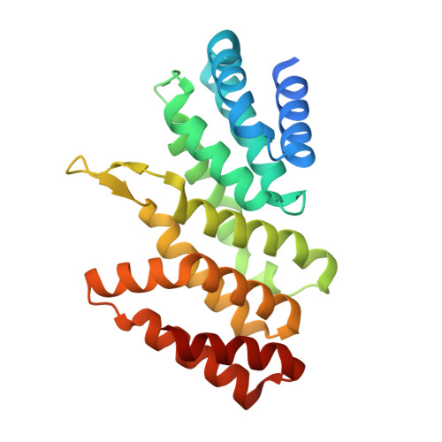
    In eukaryotes, the poly(A)-binding protein (PABP) is one of the important factors for initiation of messenger RNA translation. PABP activity is regulated by the PABP-interacting proteins (Paips), which include Paip1, Paip2A, and Paip2B. Human Paip1 has three different isoforms. Here, we report the crystal structure of the middle domain of Paip1 isoform 2 (Paip1M) as determined by single-wavelength anomalous dispersion phasing. The structure reveals a crescent-shaped domain consisting of 10 α-helices and two antiparallel β-strands forming a β-hairpin. The 10 α-helices are arranged as five HEAT repeats which form a double layer of α helices with a convex and a concave surface. Despite low sequence identity, the overall fold of Paip1M is similar to the middle domain of human eIF4GII and yeast eIF4GI. Moreover, the amino-acid sequence motif and the local structure of eIF4G involved in binding of eIF4A, are conserved in Paip1. The structure reported here is the first of a member of the Paip family, thereby filling a gap in our understanding of initiation of eukaryotic mRNA translation in three dimensions.


  • Organizational Affiliation
    • Institute of Biochemistry, Center for Structural and Cell Biology in Medicine, University of Lübeck, Ratzeburger Allee 160, Lübeck, Germany.

Macromolecule Content 

  • Total Structure Weight: 52.96 kDa 
  • Atom Count: 3,952 
  • Modeled Residue Count: 434 
  • Deposited Residue Count: 468 
  • Unique protein chains: 1

Macromolecules

Find similar proteins by:|  3D Structure
Entity ID: 1
MoleculeChains  Sequence LengthOrganismDetailsImage
Polyadenylate-binding protein-interacting protein 1
A, B
234Homo sapiensMutation(s): 0 
Gene Names: PAIP1
UniProt & NIH Common Fund Data Resources
Find proteins for Q9H074 (Homo sapiens)
Explore Q9H074 
Go to UniProtKB:  Q9H074
PHAROS:  Q9H074
GTEx:  ENSG00000172239 
Entity Groups
Sequence Clusters30% Identity50% Identity70% Identity90% Identity95% Identity100% Identity
UniProt GroupQ9H074
Sequence Annotations
Expand
Reference Sequence

Experimental Data & Validation

Experimental Data

  • Method: X-RAY DIFFRACTION
  • Resolution: 2.00 Å
  • R-Value Free:  0.244 (Depositor), 0.247 (DCC) 
  • R-Value Work:  0.190 (Depositor), 0.190 (DCC) 
  • R-Value Observed: 0.192 (Depositor) 
Space Group: P 1 21 1
Unit Cell:
Length ( Å )Angle ( ˚ )
a = 58.61α = 90
b = 76.05β = 96.47
c = 62.42γ = 90
Software Package:
Software NamePurpose
MOSFLMdata reduction
SCALAdata scaling
SHELXphasing
REFMACrefinement
PDB_EXTRACTdata extraction
MAR345dtbdata collection
SHELXCDphasing
SHELXEmodel building

Structure Validation

View Full Validation Report


Entry History 

Deposition Data

Revision History  (Full details and data files)

  • Version 1.0: 2011-05-18
    Type: Initial release
  • Version 1.1: 2011-07-13
    Changes: Version format compliance
  • Version 1.2: 2017-11-08
    Changes: Refinement description
  • Version 1.3: 2024-02-28
    Changes: Data collection, Database references