
 Download Mendeley
Crystal Strucrture of Biotin Protein Ligase from S. aureus
Pendini, N., Yap, M., Polyak, S., Cowieson, N., Daouda, T., Booker, G., Wallace, J., Wilce, M.C.J.To be published.
Experimental Data Snapshot
Starting Model: experimental
View more details
Entity ID: 1 | |||||
|---|---|---|---|---|---|
| Molecule | Chains | Sequence Length | Organism | Details | Image |
| Biotin-[acetyl-CoA-carboxylase] ligase | 323 | Staphylococcus aureus subsp. aureus ECT-R 2 | Mutation(s): 0 Gene Names: ECTR2_1310 EC: 6.3.4.15 | 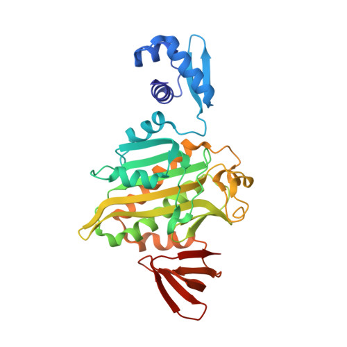 | |
UniProt | |||||
Find proteins for A0A0H3JRH8 (Staphylococcus aureus (strain Mu50 / ATCC 700699)) Explore A0A0H3JRH8 Go to UniProtKB: A0A0H3JRH8 | |||||
Entity Groups | |||||
| Sequence Clusters | 30% Identity50% Identity70% Identity90% Identity95% Identity100% Identity | ||||
| UniProt Group | A0A0H3JRH8 | ||||
Sequence AnnotationsExpand | |||||
Reference Sequence | |||||
| Ligands 1 Unique | |||||
|---|---|---|---|---|---|
| ID | Chains | Name / Formula / InChI Key | 2D Diagram | 3D Interactions | |
| BT5 Download:Ideal Coordinates CCD File | B [auth A] | BIOTINYL-5-AMP C20 H28 N7 O9 P S UTQCSTJVMLODHM-RHCAYAJFSA-N | 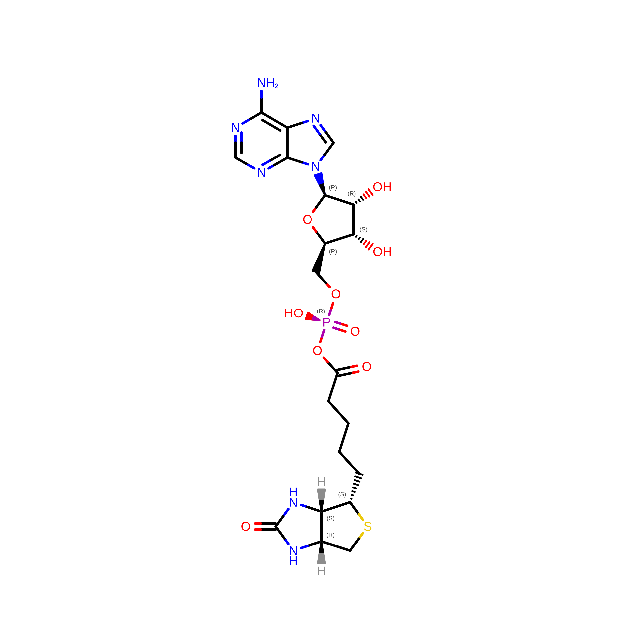 | ||
| Length ( Å ) | Angle ( ˚ ) |
|---|---|
| a = 93.564 | α = 90 |
| b = 93.564 | β = 90 |
| c = 130.65 | γ = 90 |
| Software Name | Purpose |
|---|---|
| StructureStudio | data collection |
| PHASER | phasing |
| PHENIX | refinement |
