


Crystal structure of d-mannonate dehydratase from Chromohalobacter salexigens complexed with d-arabinohydroxamate
Fedorov, A.A., Fedorov, E.V., Wichelecki, D., Gerlt, J.A., Almo, S.C.To be published.
Experimental Data Snapshot
Starting Model: experimental
View more details
Entity ID: 1 | |||||
|---|---|---|---|---|---|
| Molecule | Chains | Sequence Length | Organism | Details | Image |
| D-mannonate dehydratase | 405 | Chromohalobacter israelensis | Mutation(s): 0 Gene Names: Csal_2974 EC: 4.2.1.8 (PDB Primary Data), 4.2.1 (UniProt), 4.2.1.39 (UniProt) | 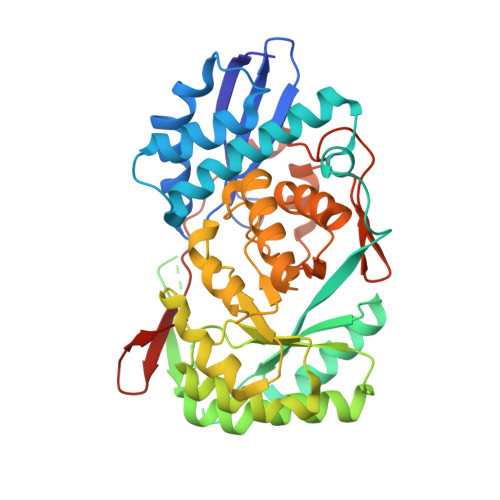 | |
UniProt | |||||
Entity Groups | |||||
| Sequence Clusters | 30% Identity50% Identity70% Identity90% Identity95% Identity100% Identity | ||||
| UniProt Group | Q1QT89 | ||||
Sequence AnnotationsExpand | |||||
Reference Sequence | |||||
| Ligands 2 Unique | |||||
|---|---|---|---|---|---|
| ID | Chains | Name / Formula / InChI Key | 2D Diagram | 3D Interactions | |
| EZ4 Download:Ideal Coordinates CCD File | F [auth A], H [auth B], J [auth C], L [auth D] | (2S,3R,4R)-2,3,4,5-tetrahydroxy-N-oxo-pentanamide C5 H9 N O6 VIMQVNVKLDVNFU-JJYYJPOSSA-N |  | ||
| CO Download:Ideal Coordinates CCD File | E [auth A], G [auth B], I [auth C], K [auth D] | COBALT (II) ION Co XLJKHNWPARRRJB-UHFFFAOYSA-N |  | ||
| Length ( Å ) | Angle ( ˚ ) |
|---|---|
| a = 110.712 | α = 90 |
| b = 179.657 | β = 90 |
| c = 85.422 | γ = 90 |
| Software Name | Purpose |
|---|---|
| ADSC | data collection |
| BALBES | phasing |
| PHENIX | refinement |
| DENZO | data reduction |
| SCALEPACK | data scaling |


