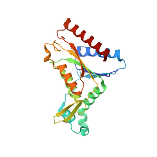
3R6W

paAzoR1 binding to nitrofurazone


Experimental Data Snapshot

  • Method: X-RAY DIFFRACTION
  • Resolution: 2.08 Å
  • R-Value Free: 0.203 
  • R-Value Work: 0.161 
  • R-Value Observed: 0.163 

Starting Model: experimental
View more details

wwPDB Validation   3D Report Full Report


Ligand Structure Quality Assessment 


This is version 1.2 of the entry. See complete history


Literature

Activation of nitrofurazone by azoreductases: multiple activities in one enzyme.

Ryan, A.Kaplan, E.Laurieri, N.Lowe, E.Sim, E.

(2011) Sci Rep 1: 63-63

  • DOI: https://doi.org/10.1038/srep00063
  • Primary Citation of Related Structures:  
    3R6W

  • PubMed Abstract: 

    Azoreductases are well known for azo pro-drug activation by gut flora. We show that azoreductases have a wider role in drug metabolism than previously thought as they can also reduce and hence activate nitrofurazone. Nitrofurazone, a nitroaromatic drug, is a broad spectrum antibiotic which has until now been considered as activated in bacteria by nitroreductases. The structure of the azoreductase with nitrofurazone bound was solved at 2.08 Å and shows nitrofurazone in an active conformation. Based on the structural information, the kinetics and stoichiometry of nitrofurazone reduction by azoreductase from P. aeruginosa, we propose a mechanism of activation which accounts for the ability of azoreductases to reduce both azo and nitroaromatic drugs. This mode of activation can explain the cytotoxic side-effects of nitrofurazone through human azoreductase homologues.


  • Organizational Affiliation

    Pharmacology Department, University of Oxford, OX1 3QT.


Macromolecules
Find similar proteins by:  (by identity cutoff)  |  3D Structure
Entity ID: 1
MoleculeChains Sequence LengthOrganismDetailsImage
FMN-dependent NADH-azoreductase 1
A, B
212Pseudomonas aeruginosaMutation(s): 0 
Gene Names: azoR1PA0785
EC: 1.7 (PDB Primary Data), 1.6.5 (UniProt), 1.7.1.17 (UniProt)
UniProt
Find proteins for Q9I5F3 (Pseudomonas aeruginosa (strain ATCC 15692 / DSM 22644 / CIP 104116 / JCM 14847 / LMG 12228 / 1C / PRS 101 / PAO1))
Explore Q9I5F3 
Go to UniProtKB:  Q9I5F3
Entity Groups  
Sequence Clusters30% Identity50% Identity70% Identity90% Identity95% Identity100% Identity
UniProt GroupQ9I5F3
Sequence Annotations
Expand
  • Reference Sequence
Small Molecules
Ligands 4 Unique
IDChains Name / Formula / InChI Key2D Diagram3D Interactions
FMN
Query on FMN

Download Ideal Coordinates CCD File 
C [auth A],
L [auth B]
FLAVIN MONONUCLEOTIDE
C17 H21 N4 O9 P
FVTCRASFADXXNN-SCRDCRAPSA-N
NFZ
Query on NFZ

Download Ideal Coordinates CCD File 
D [auth A],
E [auth A]
NITROFURAZONE
C6 H6 N4 O4
IAIWVQXQOWNYOU-FPYGCLRLSA-N
SO4
Query on SO4

Download Ideal Coordinates CCD File 
F [auth A]SULFATE ION
O4 S
QAOWNCQODCNURD-UHFFFAOYSA-L
GOL
Query on GOL

Download Ideal Coordinates CCD File 
G [auth A]
H [auth A]
I [auth A]
J [auth A]
K [auth A]
G [auth A],
H [auth A],
I [auth A],
J [auth A],
K [auth A],
M [auth B],
N [auth B]
GLYCEROL
C3 H8 O3
PEDCQBHIVMGVHV-UHFFFAOYSA-N
Experimental Data & Validation

Experimental Data

  • Method: X-RAY DIFFRACTION
  • Resolution: 2.08 Å
  • R-Value Free: 0.203 
  • R-Value Work: 0.161 
  • R-Value Observed: 0.163 
  • Space Group: P 31 2 1
Unit Cell:
Length ( Å )Angle ( ˚ )
a = 82.202α = 90
b = 82.202β = 90
c = 108.462γ = 120
Software Package:
Software NamePurpose
SCALAdata scaling
PHASERphasing
PHENIXrefinement
PDB_EXTRACTdata extraction
XDSdata scaling
XDSdata reduction

Structure Validation

View Full Validation Report


Ligand Structure Quality Assessment 


Entry History 

Deposition Data

Revision History  (Full details and data files)

  • Version 1.0: 2011-08-24
    Type: Initial release
  • Version 1.1: 2012-03-07
    Changes: Database references
  • Version 1.2: 2023-09-13
    Changes: Data collection, Database references, Derived calculations, Refinement description