3R4Z | pdb_00003r4z

Crystal structure of alpha-neoagarobiose hydrolase (ALPHA-NABH) in complex with alpha-d-galactopyranose from Saccharophagus degradans 2-40


Experimental Data Snapshot

  • Method: X-RAY DIFFRACTION
  • Resolution: 1.55 Å
  • R-Value Free: 
    0.185 (Depositor), 0.186 (DCC) 
  • R-Value Work: 
    0.164 (Depositor), 0.165 (DCC) 
  • R-Value Observed: 
    0.165 (Depositor) 

wwPDB Validation 3D Report Full Report

Validation slider image for 3R4Z

Ligand Structure Quality Assessment 


This is version 1.2 of the entry. See complete history

Literature

Crystal structure of a key enzyme in the agarolytic pathway, alpha-neoagarobiose hydrolase from Saccharophagus degradans 2-40

Ha, S.C.Lee, S.Lee, J.Kim, H.T.Ko, H.J.Kim, K.H.Choi, I.G.

(2011) Biochem Biophys Res Commun 412: 238-244

  • DOI: https://doi.org/10.1016/j.bbrc.2011.07.073
  • Primary Citation Related Structures: 
    3R4Y, 3R4Z

  • PubMed Abstract: 

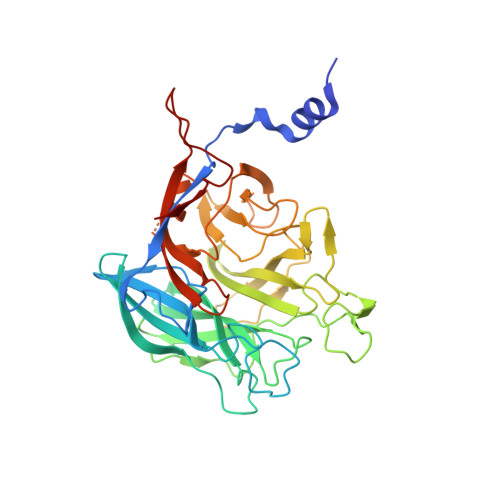
    In agarolytic microorganisms, α-neoagarobiose hydrolase (NABH) is an essential enzyme to metabolize agar because it converts α-neoagarobiose (O-3,6-anhydro-alpha-l-galactopyranosyl-(1,3)-d-galactose) into fermentable monosaccharides (d-galactose and 3,6-anhydro-l-galactose) in the agarolytic pathway. NABH can be divided into two biological classes by its cellular location. Here, we describe a structure and function of cytosolic NABH from Saccharophagus degradans 2-40 in a native protein and d-galactose complex determined at 2.0 and 1.55 Å, respectively. The overall fold is organized in an N-terminal helical extension and a C-terminal five-bladed β-propeller catalytic domain. The structure of the enzyme-ligand (d-galactose) complex predicts a +1 subsite in the substrate binding pocket. The structural features may provide insights for the evolution and classification of NABH in agarolytic pathways.


  • Organizational Affiliation
    • Physical Bioscience Division, Lawrence Berkeley National Laboratory, Berkeley, CA 94720, USA.

Macromolecule Content 

  • Total Structure Weight: 85.24 kDa 
  • Atom Count: 6,467 
  • Modeled Residue Count: 717 
  • Deposited Residue Count: 748 
  • Unique protein chains: 1

Macromolecules

Find similar proteins by:|  3D Structure
Entity ID: 1
MoleculeChains  Sequence LengthOrganismDetailsImage
Glycosyl hydrolase family 32, N terminal
A, B
374Saccharophagus degradans 2-40Mutation(s): 0 
Gene Names: Sde_2657
EC: 3.2.1
UniProt
Find proteins for Q21HB2 (Saccharophagus degradans (strain 2-40 / ATCC 43961 / DSM 17024))
Explore Q21HB2 
Go to UniProtKB:  Q21HB2
Entity Groups
Sequence Clusters30% Identity50% Identity70% Identity90% Identity95% Identity100% Identity
UniProt GroupQ21HB2
Sequence Annotations
Expand
Reference Sequence

Experimental Data & Validation

Experimental Data

  • Method: X-RAY DIFFRACTION
  • Resolution: 1.55 Å
  • R-Value Free:  0.185 (Depositor), 0.186 (DCC) 
  • R-Value Work:  0.164 (Depositor), 0.165 (DCC) 
  • R-Value Observed: 0.165 (Depositor) 
Space Group: C 1 2 1
Unit Cell:
Length ( Å )Angle ( ˚ )
a = 130.207α = 90
b = 77.128β = 101.95
c = 90.971γ = 90
Software Package:
Software NamePurpose
ADSCdata collection
MOLREPphasing
REFMACrefinement
HKL-2000data reduction
HKL-2000data scaling

Structure Validation

View Full Validation Report


Ligand Structure Quality Assessment 


Entry History 

Deposition Data

Revision History  (Full details and data files)

  • Version 1.0: 2012-02-01
    Type: Initial release
  • Version 1.1: 2020-07-29
    Type: Remediation
    Reason: Carbohydrate remediation
    Changes: Data collection, Database references, Derived calculations, Structure summary
  • Version 1.2: 2024-03-20
    Changes: Data collection, Database references, Structure summary