The Catalytic Mechanism of the Hotdog-fold Enzyme Superfamily 4-Hydroxybenzoyl-CoA Thioesterase from Arthrobacter sp. Strain SU.
Song, F., Thoden, J.B., Zhuang, Z., Latham, J., Trujillo, M., Holden, H.M., Dunaway-Mariano, D.(2012) Biochemistry 51: 7000-7016
- PubMed: 22873756
- DOI: https://doi.org/10.1021/bi301059m
- Primary Citation of Related Structures:
3R32, 3R34, 3R35, 3R36, 3R37, 3R3A, 3R3B, 3R3C, 3R3D, 3R3F, 3TEA - PubMed Abstract:
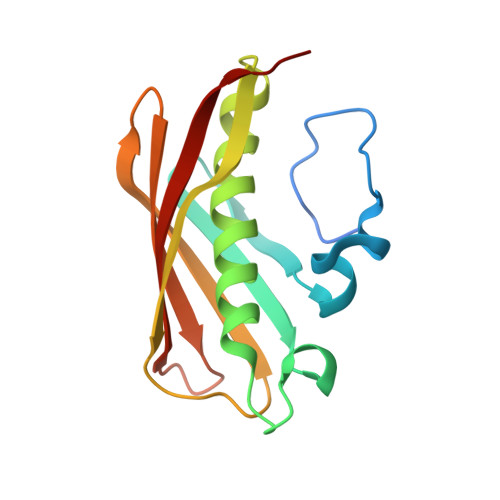
The hotdog-fold enzyme 4-hydroxybenzoyl-coenzyme A (4-HB-CoA) thioesterase from Arthrobacter sp. strain AU catalyzes the hydrolysis of 4-HB-CoA to form 4-hydroxybenzoate (4-HB) and coenzyme A (CoA) in the final step of the 4-chlorobenzoate dehalogenation pathway. Guided by the published X-ray structures of the liganded enzyme (Thoden, J. B., Zhuang, Z., Dunaway-Mariano, D., and Holden H. M. (2003) J. Biol. Chem. 278, 43709-43716), a series of site-directed mutants were prepared for testing the roles of active site residues in substrate binding and catalysis. The mutant thioesterases were subjected to X-ray structure determination to confirm retention of the native fold, and in some cases, to reveal changes in the active site configuration. In parallel, the wild-type and mutant thioesterases were subjected to transient and steady-state kinetic analysis, and to (18)O-solvent labeling experiments. Evidence is provided that suggests that Glu73 functions in nucleophilic catalysis, that Gly65 and Gln58 contribute to transition-state stabilization via hydrogen bond formation with the thioester moiety and that Thr77 orients the water nucleophile for attack at the 4-hydroxybenzoyl carbon of the enzyme-anhydride intermediate. The replacement of Glu73 with Asp was shown to switch the function of the carboxylate residue from nucleophilic catalysis to base catalysis and thus, the reaction from a two-step process involving a covalent enzyme intermediate to a single-step hydrolysis reaction. The E73D/T77A double mutant regained most of the catalytic efficiency lost in the E73D single mutant. The results from (31)P NMR experiments indicate that the substrate nucleotide unit is bound to the enzyme surface. Kinetic analysis of site-directed mutants was carried out to determine the contributions made by Arg102, Arg150, Ser120, and Thr121 in binding the nucleotide unit. Lastly, we show by kinetic and X-ray analyses of Asp31, His64, and Glu78 site-directed mutants that these three active site residues are important for productive binding of the substrate 4-hydroxybenzoyl ring.
Organizational Affiliation:
Department of Chemistry and Chemical Biology, University of New Mexico, Albuquerque, NM 87131, USA.


