


Crystal structure of enolase superfamily member from VIBRIONALES BACTERIUM complexed with Mg and Glycerol in the active site
Fedorov, A.A., Fedorov, E.V., Wichelecki, D., Gerlt, J.A., Almo, S.C.To be published.
Experimental Data Snapshot
wwPDB Validation 3D Report Full Report

Macromolecule Content
Entity ID: 1 | |||||
|---|---|---|---|---|---|
| Molecule | Chains | Sequence Length | Organism | Details | Image |
| mandelate racemase / muconate lactonizing enzyme | 401 | Vibrionales bacterium SWAT-3 | Mutation(s): 0 Gene Names: VSWAT3_13707 | 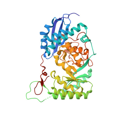 | |
UniProt | |||||
Entity Groups | |||||
| Sequence Clusters | 30% Identity50% Identity70% Identity90% Identity95% Identity100% Identity | ||||
| UniProt Group | A5KUH4 | ||||
Sequence AnnotationsExpand | |||||
Reference Sequence | |||||
| Ligands 2 Unique | |||||
|---|---|---|---|---|---|
| ID | Chains | Name / Formula / InChI Key | 2D Diagram | 3D Interactions | |
| GOL Download:Ideal Coordinates CCD File | J [auth A] M [auth B] O [auth C] R [auth D] T [auth E] | GLYCEROL C3 H8 O3 PEDCQBHIVMGVHV-UHFFFAOYSA-N |  | ||
| MG Download:Ideal Coordinates CCD File | I [auth A] K [auth B] L [auth B] N [auth C] P [auth D] | MAGNESIUM ION Mg JLVVSXFLKOJNIY-UHFFFAOYSA-N |  | ||
| Length ( Å ) | Angle ( ˚ ) |
|---|---|
| a = 167.986 | α = 90 |
| b = 211.516 | β = 90 |
| c = 211.672 | γ = 90 |
| Software Name | Purpose |
|---|---|
| ADSC | data collection |
| BALBES | phasing |
| PHENIX | refinement |
| DENZO | data reduction |
| SCALEPACK | data scaling |


