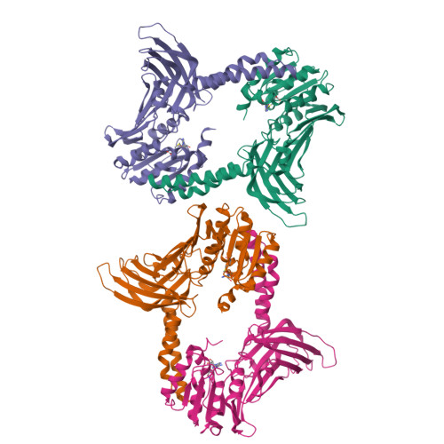
3R0Q

A Uniquely Open Conformation Revealed in the Crystal Structure of Arabidopsis Thaliana Protein Arginine Methyltransferase 10


Experimental Data Snapshot

  • Method: X-RAY DIFFRACTION
  • Resolution: 2.61 Å
  • R-Value Free: 0.224 
  • R-Value Work: 0.182 
  • R-Value Observed: 0.185 

wwPDB Validation   3D Report Full Report


Ligand Structure Quality Assessment 

Created with Raphaël 2.3.0Worse 01 BetterLigand structure goodness of fit to experimental dataBest fitted SAHClick on this verticalbar to view details

This is version 1.3 of the entry. See complete history


Literature

Crystal structure of the plant epigenetic protein arginine methyltransferase 10.

Cheng, Y.Frazier, M.Lu, F.Cao, X.Redinbo, M.R.

(2011) J Mol Biol 414: 106-122

  • DOI: https://doi.org/10.1016/j.jmb.2011.09.040
  • Primary Citation of Related Structures:  
    3R0Q

  • PubMed Abstract: 

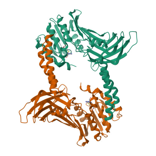
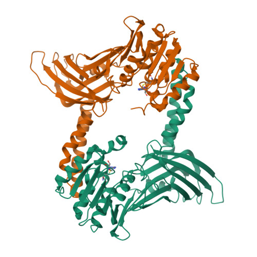
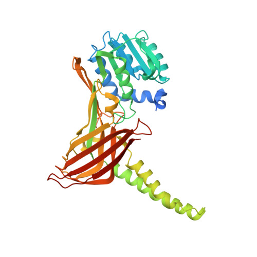
    Protein arginine methyltransferase 10 (PRMT10) is a type I arginine methyltransferase that is essential for regulating flowering time in Arabidopsis thaliana. We present a 2.6 Å resolution crystal structure of A. thaliana PRMT 10 (AtPRMT10) in complex with a reaction product, S-adenosylhomocysteine. The structure reveals a dimerization arm that is 12-20 residues longer than PRMT structures elucidated previously; as a result, the essential AtPRMT10 dimer exhibits a large central cavity and a distinctly accessible active site. We employ molecular dynamics to examine how dimerization facilitates AtPRMT10 motions necessary for activity, and we show that these motions are conserved in other PRMT enzymes. Finally, functional data reveal that the 10 N-terminal residues of AtPRMT10 influence substrate specificity, and that enzyme activity is dependent on substrate protein sequences distal from the methylation site. Taken together, these data provide insights into the molecular mechanism of AtPRMT10, as well as other members of the PRMT family of enzymes. They highlight differences between AtPRMT10 and other PRMTs but also indicate that motions are a conserved element of PRMT function.


  • Organizational Affiliation

    Department of Biochemistry and Biophysics, University of North Carolina at Chapel Hill, Chapel Hill, NC 27599, USA.


Macromolecules
Find similar proteins by:  (by identity cutoff)  |  3D Structure
Entity ID: 1
MoleculeChains Sequence LengthOrganismDetailsImage
Probable protein arginine N-methyltransferase 4.2A [auth C],
B [auth G],
C [auth A],
D [auth E]
376Arabidopsis thalianaMutation(s): 0 
Gene Names: PRMT4.2At1g04870F13M7.14F13M7_12
EC: 2.1.1 (PDB Primary Data), 2.1.1.319 (UniProt)
UniProt
Find proteins for Q9MAT5 (Arabidopsis thaliana)
Explore Q9MAT5 
Go to UniProtKB:  Q9MAT5
Entity Groups  
Sequence Clusters30% Identity50% Identity70% Identity90% Identity95% Identity100% Identity
UniProt GroupQ9MAT5
Sequence Annotations
Expand
  • Reference Sequence
Experimental Data & Validation

Experimental Data

  • Method: X-RAY DIFFRACTION
  • Resolution: 2.61 Å
  • R-Value Free: 0.224 
  • R-Value Work: 0.182 
  • R-Value Observed: 0.185 
  • Space Group: P 1 21 1
Unit Cell:
Length ( Å )Angle ( ˚ )
a = 80.552α = 90
b = 86.691β = 89.97
c = 114.735γ = 90
Software Package:
Software NamePurpose
HKL-2000data collection
PHENIXmodel building
PHENIXrefinement
HKL-2000data reduction
HKL-2000data scaling
PHENIXphasing

Structure Validation

View Full Validation Report


Ligand Structure Quality Assessment 

Created with Raphaël 2.3.0Worse 01 BetterLigand structure goodness of fit to experimental dataBest fitted SAHClick on this verticalbar to view details

Entry History 

Deposition Data

Revision History  (Full details and data files)

  • Version 1.0: 2011-10-19
    Type: Initial release
  • Version 1.1: 2011-10-26
    Changes: Database references
  • Version 1.2: 2011-12-14
    Changes: Database references
  • Version 1.3: 2024-02-21
    Changes: Data collection, Database references, Derived calculations