3QXC | pdb_00003qxc

Crystal structure of dethiobiotin synthetase (BioD) from Helicobacter pylori complexed with ATP


Experimental Data Snapshot

  • Method: X-RAY DIFFRACTION
  • Resolution: 1.34 Å
  • R-Value Free: 
    0.173 (Depositor), 0.186 (DCC) 
  • R-Value Work: 
    0.138 (Depositor) 
  • R-Value Observed: 
    0.140 (Depositor) 

Starting Model: experimental
View more details

wwPDB Validation 3D Report Full Report

Validation slider image for 3QXC

Ligand Structure Quality Assessment 


This is version 1.5 of the entry. See complete history

Literature

Structural characterization of Helicobacter pylori dethiobiotin synthetase reveals differences between family members.

Porebski, P.J.Klimecka, M.Chruszcz, M.Nicholls, R.A.Murzyn, K.Cuff, M.E.Xu, X.Cymborowski, M.Murshudov, G.N.Savchenko, A.Edwards, A.Minor, W.

(2012) FEBS J 279: 1093-1105

  • DOI: https://doi.org/10.1111/j.1742-4658.2012.08506.x
  • Primary Citation Related Structures: 
    2QMO, 3MLE, 3QXC, 3QXH, 3QXJ, 3QXS, 3QXX, 3QY0

  • PubMed Abstract: 

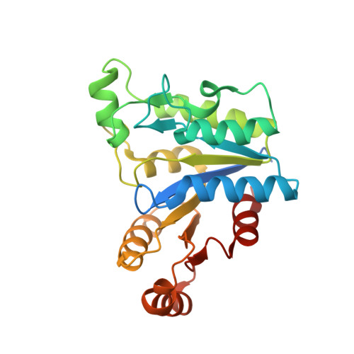
    Dethiobiotin synthetase (DTBS) is involved in the biosynthesis of biotin in bacteria, fungi, and plants. As humans lack this pathway, DTBS is a promising antimicrobial drug target. We determined structures of DTBS from Helicobacter pylori (hpDTBS) bound with cofactors and a substrate analog, and described its unique characteristics relative to other DTBS proteins. Comparison with bacterial DTBS orthologs revealed considerable structural differences in nucleotide recognition. The C-terminal region of DTBS proteins, which contains two nucleotide-recognition motifs, differs greatly among DTBS proteins from different species. The structure of hpDTBS revealed that this protein is unique and does not contain a C-terminal region containing one of the motifs. The single nucleotide-binding motif in hpDTBS is similar to its counterpart in GTPases; however, isothermal titration calorimetry binding studies showed that hpDTBS has a strong preference for ATP. The structural determinants of ATP specificity were assessed with X-ray crystallographic studies of hpDTBS·ATP and hpDTBS·GTP complexes. The unique mode of nucleotide recognition in hpDTBS makes this protein a good target for H. pylori-specific inhibitors of the biotin synthesis pathway.


  • Organizational Affiliation
    • Department of Molecular Physiology and Biological Physics, University of Virginia, Charlottesville, VA, USA.

Macromolecule Content 

  • Total Structure Weight: 28.68 kDa 
  • Atom Count: 2,317 
  • Modeled Residue Count: 224 
  • Deposited Residue Count: 242 
  • Unique protein chains: 1

Macromolecules

Find similar proteins by:|  3D Structure
Entity ID: 1
MoleculeChains  Sequence LengthOrganismDetailsImage
Dethiobiotin synthetase242Helicobacter pylori 26695Mutation(s): 0 
Gene Names: bioDhp0029HP_0029
EC: 6.3.3.3
UniProt
Find proteins for O24872 (Helicobacter pylori (strain ATCC 700392 / 26695))
Explore O24872 
Go to UniProtKB:  O24872
Entity Groups
Sequence Clusters30% Identity50% Identity70% Identity90% Identity95% Identity100% Identity
UniProt GroupO24872
Sequence Annotations
Expand
Reference Sequence

Small Molecules

Ligands 6 Unique
IDChains Name / Formula / InChI Key2D Diagram3D Interactions
ATP

Query on ATP


Download:Ideal Coordinates CCD File
B [auth A]ADENOSINE-5'-TRIPHOSPHATE
C10 H16 N5 O13 P3
ZKHQWZAMYRWXGA-KQYNXXCUSA-N
PEG

Query on PEG


Download:Ideal Coordinates CCD File
Q [auth A]DI(HYDROXYETHYL)ETHER
C4 H10 O3
MTHSVFCYNBDYFN-UHFFFAOYSA-N
GOL

Query on GOL


Download:Ideal Coordinates CCD File
N [auth A]GLYCEROL
C3 H8 O3
PEDCQBHIVMGVHV-UHFFFAOYSA-N
EDO

Query on EDO


Download:Ideal Coordinates CCD File
E [auth A]
F [auth A]
G [auth A]
H [auth A]
I [auth A]
E [auth A],
F [auth A],
G [auth A],
H [auth A],
I [auth A],
J [auth A],
K [auth A],
L [auth A],
M [auth A],
O [auth A],
P [auth A],
R [auth A]
1,2-ETHANEDIOL
C2 H6 O2
LYCAIKOWRPUZTN-UHFFFAOYSA-N
NO3

Query on NO3


Download:Ideal Coordinates CCD File
D [auth A]NITRATE ION
N O3
NHNBFGGVMKEFGY-UHFFFAOYSA-N
MG

Query on MG


Download:Ideal Coordinates CCD File
C [auth A]MAGNESIUM ION
Mg
JLVVSXFLKOJNIY-UHFFFAOYSA-N

Experimental Data & Validation

Experimental Data

  • Method: X-RAY DIFFRACTION
  • Resolution: 1.34 Å
  • R-Value Free:  0.173 (Depositor), 0.186 (DCC) 
  • R-Value Work:  0.138 (Depositor) 
  • R-Value Observed: 0.140 (Depositor) 
Space Group: C 1 2 1
Unit Cell:
Length ( Å )Angle ( ˚ )
a = 81.883α = 90
b = 37.691β = 101.24
c = 69.184γ = 90
Software Package:
Software NamePurpose
HKL-3000data collection
HKL-3000phasing
MOLREPphasing
REFMACrefinement
Cootmodel building
HKL-3000data reduction
HKL-3000data scaling

Structure Validation

View Full Validation Report


Ligand Structure Quality Assessment 


Entry History 

Revision History  (Full details and data files)

  • Version 1.0: 2011-03-30
    Type: Initial release
  • Version 1.1: 2011-07-13
    Changes: Version format compliance
  • Version 1.2: 2012-03-28
    Changes: Database references
  • Version 1.3: 2013-03-27
    Changes: Database references
  • Version 1.4: 2022-04-13
    Changes: Database references, Derived calculations, Structure summary
  • Version 1.5: 2023-09-13
    Changes: Data collection, Refinement description