3QWX | pdb_00003qwx

CED-2 1-174


Experimental Data Snapshot

  • Method: X-RAY DIFFRACTION
  • Resolution: 2.01 Å
  • R-Value Free: 
    0.266 (Depositor), 0.235 (DCC) 
  • R-Value Work: 
    0.214 (Depositor) 
  • R-Value Observed: 
    0.217 (Depositor) 

wwPDB Validation 3D Report Full Report

Validation slider image for 3QWX

This is version 1.3 of the entry. See complete history

Literature

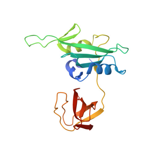
Crystal structure of the cell corpse engulfment protein CED-2 in Caenorhabditis elegans.

Kang, Y.Xu, J.Liu, Y.Sun, J.Sun, D.Hu, Y.Liu, Y.

(2011) Biochem Biophys Res Commun 410: 189-194

  • DOI: https://doi.org/10.1016/j.bbrc.2011.05.051
  • Primary Citation Related Structures: 
    3QWX, 3QWY

  • PubMed Abstract: 

    In the nematode Caenorhabditis elegans, the cell corpse engulfment proteins CED-2, CED-5, and CED-12 act in the same pathway to regulate the activation of the Rac small GTPase, CED-10, leading to the rearrangement of the actin cytoskeleton for engulfing apoptotic cells. Nevertheless, it is not well understood how these proteins act together. Here we report the crystal structures of the CED-2 protein as determined by X-ray crystallography. The full-length CED-2 protein and its truncated form containing the N-terminal SH2 domain and the first SH3 domain show similar three-dimensional structures. A CED-2 point mutation (F125G) disrupting its interaction with the PXXP motif of CED-5 did not affect its rescuing activity. However, CED-2 was found to interact with the N-terminal region of CED-5. Our findings suggest that CED-2 may regulate cell corpse engulfment by interacting with CED-5 through the N-terminal region rather than the PXXP motif.


  • Organizational Affiliation
    • National Laboratory of Biomacromolecules, Institute of Biophysics, Chinese Academy of Sciences, Beijing 100101, China.

Macromolecule Content 

  • Total Structure Weight: 19.66 kDa 
  • Atom Count: 1,471 
  • Modeled Residue Count: 171 
  • Deposited Residue Count: 174 
  • Unique protein chains: 1

Macromolecules

Find similar proteins by:|  3D Structure
Entity ID: 1
MoleculeChains  Sequence LengthOrganismDetailsImage
Cell death abnormality protein 2A [auth X]174Caenorhabditis elegansMutation(s): 0 
Gene Names: ced-2Y41D4B.13
UniProt
Find proteins for Q9NHC3 (Caenorhabditis elegans)
Explore Q9NHC3 
Go to UniProtKB:  Q9NHC3
Entity Groups
Sequence Clusters30% Identity50% Identity70% Identity90% Identity95% Identity100% Identity
UniProt GroupQ9NHC3
Sequence Annotations
Expand
Reference Sequence

Small Molecules

Ligands 1 Unique
IDChains Name / Formula / InChI Key2D Diagram3D Interactions
SO4

Query on SO4


Download:Ideal Coordinates CCD File
B [auth X]SULFATE ION
O4 S
QAOWNCQODCNURD-UHFFFAOYSA-L

Experimental Data & Validation

Experimental Data

  • Method: X-RAY DIFFRACTION
  • Resolution: 2.01 Å
  • R-Value Free:  0.266 (Depositor), 0.235 (DCC) 
  • R-Value Work:  0.214 (Depositor) 
  • R-Value Observed: 0.217 (Depositor) 
Space Group: P 61 2 2
Unit Cell:
Length ( Å )Angle ( ˚ )
a = 86.681α = 90
b = 86.681β = 90
c = 87.781γ = 120
Software Package:
Software NamePurpose
CrystalCleardata collection
SOLVEphasing
REFMACrefinement
HKL-2000data reduction
HKL-2000data scaling

Structure Validation

View Full Validation Report


Entry History 

Deposition Data

Revision History  (Full details and data files)

  • Version 1.0: 2011-06-08
    Type: Initial release
  • Version 1.1: 2011-07-13
    Changes: Version format compliance
  • Version 1.2: 2011-07-20
    Changes: Database references
  • Version 1.3: 2024-02-21
    Changes: Data collection, Database references, Derived calculations