3QUK

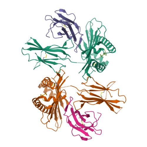
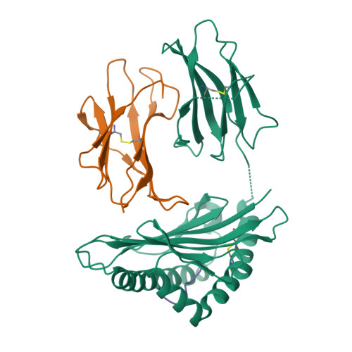
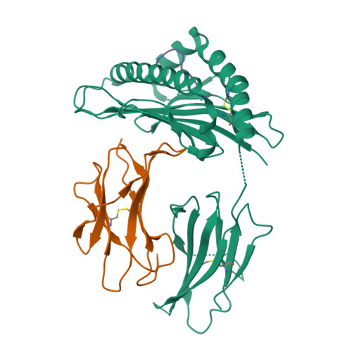
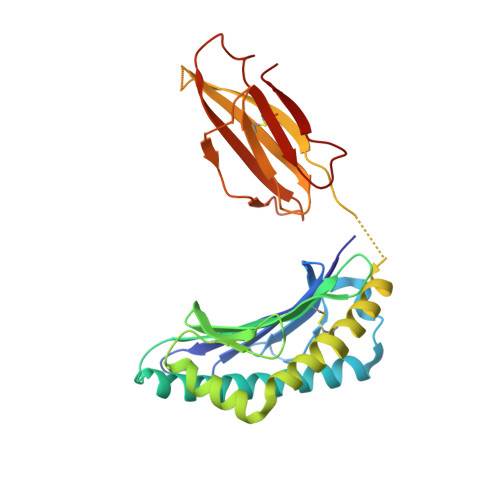
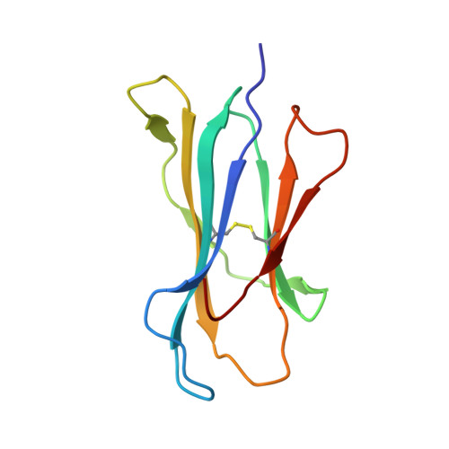
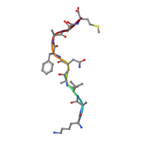
Crystal structures of the murine class I major histocompatibility complex H-2Db in complex with LCMV-derived gp33 altered peptide ligand (Y4A)


Experimental Data Snapshot

  • Method: X-RAY DIFFRACTION
  • Resolution: 2.41 Å
  • R-Value Free: 0.285 
  • R-Value Work: 0.242 
  • R-Value Observed: 0.244 

Starting Model: experimental
View more details

wwPDB Validation   3D Report Full Report


This is version 1.5 of the entry. See complete history


Literature

Unexpected T-cell recognition of an altered peptide ligand is driven by reversed thermodynamics.

Allerbring, E.B.Duru, A.D.Uchtenhagen, H.Madhurantakam, C.Tomek, M.B.Grimm, S.Mazumdar, P.A.Friemann, R.Uhlin, M.Sandalova, T.Nygren, P.A.Achour, A.

(2012) Eur J Immunol 42: 2990-3000

  • DOI: https://doi.org/10.1002/eji.201242588
  • Primary Citation of Related Structures:  
    3QUK, 3QUL

  • PubMed Abstract: 

    The molecular basis underlying T-cell recognition of MHC molecules presenting altered peptide ligands is still not well-established. A hierarchy of T-cell activation by MHC class I-restricted altered peptide ligands has been defined using the T-cell receptor P14 specific for H-2D(b) in complex with the immunodominant lymphocytic choriomeningitis virus peptide gp33 (KAVYNFATM). While substitution of tyrosine to phenylalanine (Y4F) or serine (Y4S) abolished recognition by P14, the TCR unexpectedly recognized H-2D(b) in complex with the alanine-substituted semiagonist Y4A, which displayed the most significant structural modification. The observed functional hierarchy gp33 > Y4A > Y4S = Y4F was neither due to higher stabilization capacity nor to differences in structural conformation. However, thermodynamic analysis demonstrated that while recognition of the full agonist H-2D(b) /gp33 was strictly enthalpy driven, recognition of the weak agonist H-2D(b) /Y4A was instead entropy driven with a large reduction in the favorable enthalpy term. The fourfold larger negative heat capacity derived for the interaction of P14 with H-2D(b) /gp33 compared with H-2D(b) /Y4A can possibly be explained by higher water entrapment at the TCR/MHC interface, which is also consistent with the measured opposite entropy contributions for the interactions of P14 with both MHCs. In conclusion, this study demonstrates that P14 makes use of different strategies to adapt to structural modifications in the MHC/peptide complex.


  • Organizational Affiliation

    Center for Infectious Medicine, Department of Medicine, Karolinska University Hospital Huddinge, Karolinska Institutet, Stockholm, Sweden.


Macromolecules
Find similar proteins by:  (by identity cutoff)  |  3D Structure
Entity ID: 1
MoleculeChains Sequence LengthOrganismDetailsImage
H-2 class I histocompatibility antigen, D-B alpha chain
A, D
276Mus musculusMutation(s): 0 
Gene Names: H2-D1
UniProt
Find proteins for P01899 (Mus musculus)
Explore P01899 
Go to UniProtKB:  P01899
Entity Groups  
Sequence Clusters30% Identity50% Identity70% Identity90% Identity95% Identity100% Identity
UniProt GroupP01899
Sequence Annotations
Expand
  • Reference Sequence
Find similar proteins by:  (by identity cutoff)  |  3D Structure
Entity ID: 2
MoleculeChains Sequence LengthOrganismDetailsImage
Beta-2-microglobulin
B, E
99Mus musculusMutation(s): 1 
Gene Names: B2m
UniProt & NIH Common Fund Data Resources
Find proteins for P01887 (Mus musculus)
Explore P01887 
Go to UniProtKB:  P01887
IMPC:  MGI:88127
Entity Groups  
Sequence Clusters30% Identity50% Identity70% Identity90% Identity95% Identity100% Identity
UniProt GroupP01887
Sequence Annotations
Expand
  • Reference Sequence

Find similar proteins by:  Sequence   |   3D Structure  

Entity ID: 3
MoleculeChains Sequence LengthOrganismDetailsImage
Pre-glycoprotein polyprotein GP complex
C, F
9Lymphocytic choriomeningitis virus (strain WE)Mutation(s): 2 
UniProt
Find proteins for P07399 (Lymphocytic choriomeningitis virus (strain WE))
Explore P07399 
Go to UniProtKB:  P07399
Entity Groups  
UniProt GroupP07399
Sequence Annotations
Expand
  • Reference Sequence
Experimental Data & Validation

Experimental Data

  • Method: X-RAY DIFFRACTION
  • Resolution: 2.41 Å
  • R-Value Free: 0.285 
  • R-Value Work: 0.242 
  • R-Value Observed: 0.244 
  • Space Group: C 1 2 1
Unit Cell:
Length ( Å )Angle ( ˚ )
a = 118.51α = 90
b = 126.21β = 124.54
c = 96.47γ = 90
Software Package:
Software NamePurpose
HKL-2000data collection
PHASERphasing
REFMACrefinement
MOSFLMdata reduction
SCALAdata scaling

Structure Validation

View Full Validation Report


Entry History 

Revision History  (Full details and data files)

  • Version 1.0: 2012-03-21
    Type: Initial release
  • Version 1.1: 2017-04-12
    Changes: Database references
  • Version 1.2: 2017-05-03
    Changes: Structure summary
  • Version 1.3: 2018-03-07
    Changes: Data collection
  • Version 1.4: 2023-09-13
    Changes: Data collection, Database references, Derived calculations, Refinement description
  • Version 1.5: 2024-10-16
    Changes: Structure summary