3QTK

The crystal structure of chemically synthesized VEGF-A


Experimental Data Snapshot

  • Method: X-RAY DIFFRACTION
  • Resolution: 1.85 Å
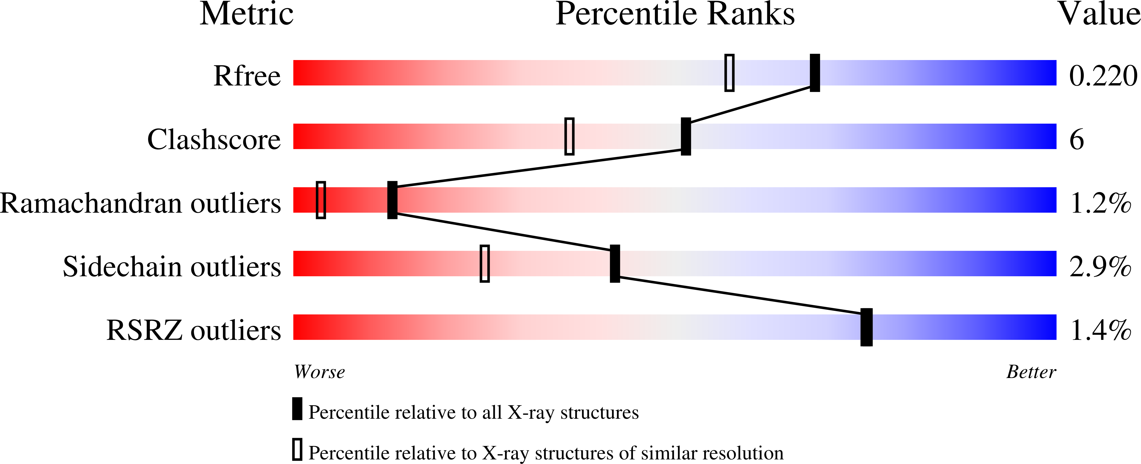
  • R-Value Free: 0.223 
  • R-Value Work: 0.180 
  • R-Value Observed: 0.182 

Starting Model: experimental
View more details

wwPDB Validation   3D Report Full Report


This is version 1.3 of the entry. See complete history


Literature

Total chemical synthesis of biologically active vascular endothelial growth factor.

Mandal, K.Kent, S.B.

(2011) Angew Chem Int Ed Engl 50: 8029-8033

  • DOI: https://doi.org/10.1002/anie.201103237
  • Primary Citation of Related Structures:  
    3QTK

  • PubMed Abstract: 

    Efficient access: the 204-residue covalent-dimer vascular endothelial growth factor with full mitogenic activity was prepared from three unprotected peptide segments by one-pot native chemical ligations. The covalent structure of the synthetic VEGF was confirmed by precise mass measurement, and the three-dimensional structure of the synthetic protein was determined by high-resolution X-ray crystallography.


  • Organizational Affiliation

    Department of Biochemistry and Molecular Biology, Institute for Biophysical Dynamics, The University of Chicago, IL 60637, USA.


Macromolecules
Find similar proteins by:  (by identity cutoff)  |  3D Structure
Entity ID: 1
MoleculeChains Sequence LengthOrganismDetailsImage
Vascular endothelial growth factor A102Homo sapiensMutation(s): 0 
UniProt & NIH Common Fund Data Resources
Find proteins for P15692 (Homo sapiens)
Go to UniProtKB:  P15692
PHAROS:  P15692
GTEx:  ENSG00000112715 
Sequence Annotations
Expand
  • Reference Sequence
Small Molecules
Ligands 3 Unique
IDChains Name / Formula / InChI Key2D Diagram3D Interactions
TFA
Query on TFA

Download Ideal Coordinates CCD File 
H [auth A]
J [auth A]
K [auth A]
M [auth D]
O [auth B]
H [auth A],
J [auth A],
K [auth A],
M [auth D],
O [auth B],
X [auth F]
trifluoroacetic acid
C2 H F3 O2
DTQVDTLACAAQTR-UHFFFAOYSA-N
GOL
Query on GOL

Download Ideal Coordinates CCD File 
I [auth A],
N [auth D],
R [auth C],
S [auth C]
GLYCEROL
C3 H8 O3
PEDCQBHIVMGVHV-UHFFFAOYSA-N
ACT
Query on ACT

Download Ideal Coordinates CCD File 
G [auth A]
L [auth D]
P [auth B]
Q [auth C]
T [auth C]
G [auth A],
L [auth D],
P [auth B],
Q [auth C],
T [auth C],
U [auth E],
V [auth E],
W [auth F]
ACETATE ION
C2 H3 O2
QTBSBXVTEAMEQO-UHFFFAOYSA-M
Experimental Data & Validation

Experimental Data

  • Method: X-RAY DIFFRACTION
  • Resolution: 1.85 Å
  • R-Value Free: 0.223 
  • R-Value Work: 0.180 
  • R-Value Observed: 0.182 
  • Space Group: C 1 2 1
Unit Cell:
Length ( Å )Angle ( ˚ )
a = 230.922α = 90
b = 44.008β = 99.93
c = 73.111γ = 90
Software Package:
Software NamePurpose
SCALEPACKdata scaling
PHENIXrefinement
PDB_EXTRACTdata extraction
ADSCdata collection
DENZOdata reduction
PHASERphasing

Structure Validation

View Full Validation Report


Entry History 

Deposition Data

Revision History  (Full details and data files)

  • Version 1.0: 2011-07-27
    Type: Initial release
  • Version 1.1: 2012-10-17
    Changes: Database references
  • Version 1.2: 2023-09-13
    Changes: Data collection, Database references, Derived calculations, Refinement description
  • Version 1.3: 2024-10-16
    Changes: Structure summary