3QSU | pdb_00003qsu

Structure of Staphylococcus aureus Hfq in complex with A7 RNA


Experimental Data Snapshot

  • Method: X-RAY DIFFRACTION
  • Resolution: 2.20 Å
  • R-Value Free: 
    0.255 (Depositor), 0.258 (DCC) 
  • R-Value Work: 
    0.196 (Depositor), 0.196 (DCC) 
  • R-Value Observed: 
    0.198 (Depositor) 

wwPDB Validation 3D Report Full Report

Validation slider image for 3QSU

This is version 1.5 of the entry. See complete history

Literature

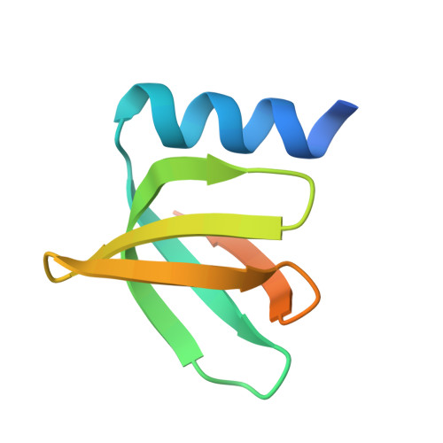
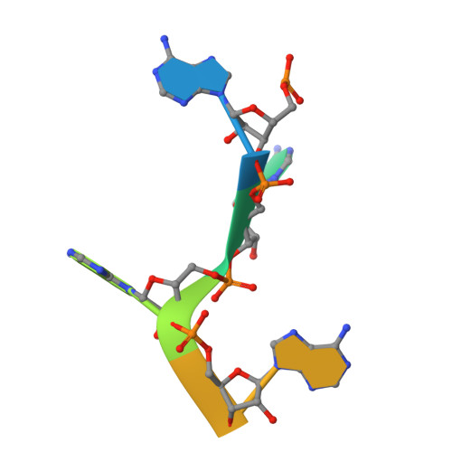
Structural mechanism of Staphylococcus aureus Hfq binding to an RNA A-tract.

Horstmann, N.Orans, J.Valentin-Hansen, P.Shelburne, S.A.Brennan, R.G.

(2012) Nucleic Acids Res 40: 11023-11035

  • DOI: https://doi.org/10.1093/nar/gks809
  • Primary Citation Related Structures: 
    3QSU

  • PubMed Abstract: 

    Hfq is a post-transcriptional regulator that plays a key role in bacterial gene expression by binding AU-rich sequences and A-tracts to facilitate the annealing of sRNAs to target mRNAs and to affect RNA stability. To understand how Hfq from the Gram-positive bacterium Staphylococcus aureus (Sa) binds A-tract RNA, we determined the crystal structure of an Sa Hfq-adenine oligoribonucleotide complex. The structure reveals a bipartite RNA-binding motif on the distal face that is composed of a purine nucleotide-specificity site (R-site) and a non-discriminating linker site (L-site). The (R-L)-binding motif, which is also utilized by Bacillus subtilis Hfq to bind (AG)(3)A, differs from the (A-R-N) tripartite poly(A) RNA-binding motif of Escherichia coli Hfq whereby the Sa Hfq R-site strongly prefers adenosine, is more aromatic and permits deeper insertion of the adenine ring. R-site adenine-stacking residue Phe30, which is conserved among Gram-positive bacterial Hfqs, and an altered conformation about β3 and β4 eliminate the adenosine-specificity site (A-site) and create the L-site. Binding studies show that Sa Hfq binds (AU)(3)A ≈ (AG)(3)A ≥ (AC)(3)A > (AA)(3)A and L-site residue Lys33 plays a significant role. The (R-L) motif is likely utilized by Hfqs from most Gram-positive bacteria to bind alternating (A-N)(n) RNA.


  • Organizational Affiliation
    • Department of Biochemistry and Molecular Biology, UT MD Anderson Cancer Center, Houston, TX 77030, USA.

Macromolecule Content 

  • Total Structure Weight: 129.43 kDa 
  • Atom Count: 7,325 
  • Modeled Residue Count: 859 
  • Deposited Residue Count: 1,092 
  • Unique protein chains: 1
  • Unique nucleic acid chains: 1

Macromolecules


Find similar proteins by:|  3D Structure
Find similar nucleic acids by:  Sequence
Entity ID: 2
MoleculeChains LengthOrganismImage
RNA (5'-R(P*AP*AP*AP*AP*AP*AP*A)-3')O [auth R],
P
7N/A
Sequence Annotations
Expand
Reference Sequence

Small Molecules

Ligands 1 Unique
IDChains Name / Formula / InChI Key2D Diagram3D Interactions
ZN

Query on ZN


Download:Ideal Coordinates CCD File
AA [auth M]
BA [auth M]
CA [auth C]
DA [auth C]
EA [auth C]
AA [auth M],
BA [auth M],
CA [auth C],
DA [auth C],
EA [auth C],
FA [auth D],
GA [auth D],
HA [auth E],
IA [auth E],
JA [auth F],
KA [auth F],
LA [auth G],
MA [auth G],
NA [auth J],
OA [auth J],
PA [auth N],
Q [auth A],
QA [auth N],
R [auth A],
RA [auth S],
S [auth B],
SA [auth S],
T [auth B],
U [auth H],
V [auth H],
W [auth I],
X [auth I],
Y [auth K],
Z [auth K]
ZINC ION
Zn
PTFCDOFLOPIGGS-UHFFFAOYSA-N

Experimental Data & Validation

Experimental Data

  • Method: X-RAY DIFFRACTION
  • Resolution: 2.20 Å
  • R-Value Free:  0.255 (Depositor), 0.258 (DCC) 
  • R-Value Work:  0.196 (Depositor), 0.196 (DCC) 
  • R-Value Observed: 0.198 (Depositor) 
Space Group: P 3
Unit Cell:
Length ( Å )Angle ( ˚ )
a = 156.397α = 90
b = 156.397β = 90
c = 34.646γ = 120
Software Package:
Software NamePurpose
ADSCdata collection
PHASERphasing
PHENIXrefinement
MOSFLMdata reduction
SCALAdata scaling

Structure Validation

View Full Validation Report


Entry History 

Deposition Data

Revision History  (Full details and data files)

  • Version 1.0: 2012-09-12
    Type: Initial release
  • Version 1.1: 2012-09-26
    Changes: Database references
  • Version 1.2: 2012-12-12
    Changes: Database references
  • Version 1.3: 2013-06-19
    Changes: Derived calculations
  • Version 1.4: 2017-11-08
    Changes: Refinement description
  • Version 1.5: 2024-02-21
    Changes: Data collection, Database references, Derived calculations