3QPZ | pdb_00003qpz

Crystal structure of the N59A mutant of the 3-deoxy-d-manno-octulosonate 8-phosphate synthase (KDO8PS) from Neisseria meningitidis


Experimental Data Snapshot

  • Method: X-RAY DIFFRACTION
  • Resolution: 1.75 Å
  • R-Value Free: 
    0.215 (Depositor), 0.228 (DCC) 
  • R-Value Work: 
    0.180 (Depositor), 0.193 (DCC) 
  • R-Value Observed: 
    0.182 (Depositor) 

Starting Model: experimental
View more details

wwPDB Validation 3D Report Full Report

Validation slider image for 3QPZ

This is version 1.3 of the entry. See complete history

Literature

Targeting the role of a key conserved motif for substrate selection and catalysis by 3-deoxy-D-manno-octulosonate 8-phosphate synthase

Allison, T.M.Hutton, R.D.Cochrane, F.C.Yeoman, J.A.Jameson, G.B.Parker, E.J.

(2011) Biochemistry 50: 3686-3695

  • DOI: https://doi.org/10.1021/bi200251f
  • Primary Citation Related Structures: 
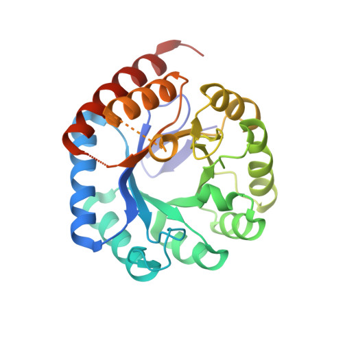
    3QPY, 3QPZ, 3QQ0, 3QQ1

  • PubMed Abstract: 

    3-Deoxy-D-manno-octulosonate 8-phosphate synthase (KDO8PS) catalyzes the reaction between three-carbon phosphoenolpyruvate (PEP) and five-carbon d-arabinose 5-phosphate (A5P), generating KDO8P, a key intermediate in the biosynthetic pathway to 3-deoxy-D-manno-octulosonate, a component of the lipopolysaccharide of the Gram-negative bacterial cell wall. Both metal-dependent and metal-independent forms of KDO8PS have been characterized. KDO8PS is evolutionarily and mechanistically related to the first enzyme of the shikimate pathway, the obligately divalent metal ion-dependent 3-deoxy-D-arabino-heptulosonate 7-phosphate synthase (DAH7PS) that couples PEP and four-carbon D-erythrose 4-phosphate (E4P) to give DAH7P. In KDO8PS, an absolutely conserved KANRS motif forms part of the A5P binding site, whereas in DAH7PS, an absolutely conserved KPR(S/T) motif accommodates E4P. Here, we have characterized four mutants of this motif (AANRS, KAARS, KARS, and KPRS) in metal-dependent KDO8PS from Acidithiobacillus ferrooxidans and metal-independent KDO8PS from Neisseria meningitidis to test the roles of the universal Lys and the Ala-Asn portion of the KANRS motif. The X-ray structures, determined for the N. meningitidis KDO8PS mutants, indicated no gross structural penalty resulting from mutation, but the subtle changes observed in the active sites of these mutant proteins correlated with their altered catalytic function. (1) The AANRS mutations destroyed catalytic activity. (2) The KAARS mutations lowered substrate selectivity, as well as activity. (3) Replacing KANRS with KARS or KPRS destroyed KDO8PS activity but did not produce a functional DAH7PS. Thus, Lys is critical to catalysis, and other changes are necessary to switch substrate specificity for both the metal-independent and metal-dependent forms of these enzymes.


  • Organizational Affiliation
    • The Riddet Institute and Institute of Fundamental Sciences, Massey University, Palmerston North, New Zealand.

Macromolecule Content 

  • Total Structure Weight: 122.49 kDa 
  • Atom Count: 8,552 
  • Modeled Residue Count: 1,009 
  • Deposited Residue Count: 1,120 
  • Unique protein chains: 1

Macromolecules

Find similar proteins by:|  3D Structure
Entity ID: 1
MoleculeChains  Sequence LengthOrganismDetailsImage
2-dehydro-3-deoxyphosphooctonate aldolase
A, B, C, D
280Neisseria meningitidis MC58Mutation(s): 1 
Gene Names: kdsANMB1283
EC: 2.5.1.55
UniProt
Find proteins for Q9JZ55 (Neisseria meningitidis serogroup B (strain ATCC BAA-335 / MC58))
Explore Q9JZ55 
Go to UniProtKB:  Q9JZ55
Entity Groups
Sequence Clusters30% Identity50% Identity70% Identity90% Identity95% Identity100% Identity
UniProt GroupQ9JZ55
Sequence Annotations
Expand
Reference Sequence

Small Molecules

Ligands 4 Unique
IDChains Name / Formula / InChI Key2D Diagram3D Interactions
PO4

Query on PO4


Download:Ideal Coordinates CCD File
G [auth B]PHOSPHATE ION
O4 P
NBIIXXVUZAFLBC-UHFFFAOYSA-K
GOL

Query on GOL


Download:Ideal Coordinates CCD File
J [auth C],
N [auth D]
GLYCEROL
C3 H8 O3
PEDCQBHIVMGVHV-UHFFFAOYSA-N
CL

Query on CL


Download:Ideal Coordinates CCD File
E [auth A]
F [auth A]
H [auth B]
I [auth B]
K [auth C]
E [auth A],
F [auth A],
H [auth B],
I [auth B],
K [auth C],
L [auth C],
O [auth D],
P [auth D]
CHLORIDE ION
Cl
VEXZGXHMUGYJMC-UHFFFAOYSA-M
NA

Query on NA


Download:Ideal Coordinates CCD File
M [auth C]SODIUM ION
Na
FKNQFGJONOIPTF-UHFFFAOYSA-N

Experimental Data & Validation

Experimental Data

  • Method: X-RAY DIFFRACTION
  • Resolution: 1.75 Å
  • R-Value Free:  0.215 (Depositor), 0.228 (DCC) 
  • R-Value Work:  0.180 (Depositor), 0.193 (DCC) 
  • R-Value Observed: 0.182 (Depositor) 
Space Group: P 21 21 21
Unit Cell:
Length ( Å )Angle ( ˚ )
a = 81.642α = 90
b = 85.368β = 90
c = 162.624γ = 90
Software Package:
Software NamePurpose
d*TREKdata scaling
REFMACrefinement
PDB_EXTRACTdata extraction
CrystalCleardata collection
d*TREKdata reduction
MOLREPphasing

Structure Validation

View Full Validation Report


Entry History 

Deposition Data

Revision History  (Full details and data files)

  • Version 1.0: 2011-04-13
    Type: Initial release
  • Version 1.1: 2011-07-13
    Changes: Version format compliance
  • Version 1.2: 2014-03-05
    Changes: Database references
  • Version 1.3: 2023-11-01
    Changes: Data collection, Database references, Derived calculations, Refinement description