3QPO | pdb_00003qpo

Structure of PDE10-inhibitor complex


Experimental Data Snapshot

  • Method: X-RAY DIFFRACTION
  • Resolution: 1.80 Å
  • R-Value Free: 
    0.236 (Depositor), 0.243 (DCC) 
  • R-Value Work: 
    0.204 (Depositor), 0.213 (DCC) 
  • R-Value Observed: 
    0.207 (Depositor) 

wwPDB Validation 3D Report Full Report

Validation slider image for 3QPO

Ligand Structure Quality Assessment 


This is version 1.3 of the entry. See complete history

Literature

Use of Structure-Based Design to Discover a Potent, Selective, In Vivo Active Phosphodiesterase 10A Inhibitor Lead Series for the Treatment of Schizophrenia.

Helal, C.J.Kang, Z.Hou, X.Pandit, J.Chappie, T.A.Humphrey, J.M.Marr, E.S.Fennell, K.F.Chenard, L.K.Fox, C.Schmidt, C.J.Williams, R.D.Chapin, D.S.Siuciak, J.Lebel, L.Menniti, F.Cianfrogna, J.Fonseca, K.R.Nelson, F.R.O'Connor, R.Macdougall, M.McDowell, L.Liras, S.

(2011) J Med Chem 54: 4536-4547

  • DOI: https://doi.org/10.1021/jm2001508
  • Primary Citation Related Structures: 
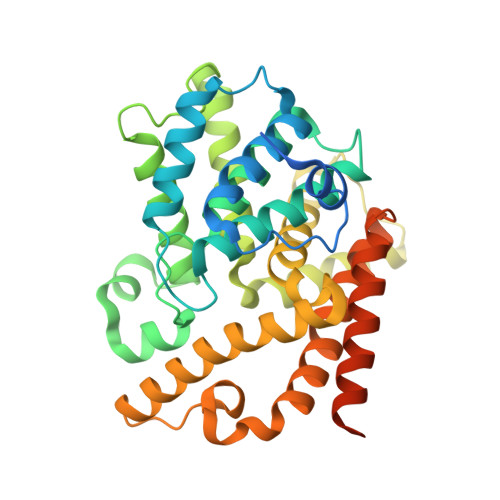
    3QPN, 3QPO, 3QPP

  • PubMed Abstract: 

    Utilizing structure-based virtual library design and scoring, a novel chimeric series of phosphodiesterase 10A (PDE10A) inhibitors was discovered by synergizing binding site interactions and ADME properties of two chemotypes. Virtual libraries were docked and scored for potential binding ability, followed by visual inspection to prioritize analogs for parallel and directed synthesis. The process yielded highly potent and selective compounds such as 16. New X-ray cocrystal structures enabled rational design of substituents that resulted in the successful optimization of physical properties to produce in vivo activity and to modulate microsomal clearance and permeability.


  • Organizational Affiliation
    • Pfizer Worldwide Research and Development, Groton, Connecticut 06340, United States. chris.j.helal@pfizer.com

Macromolecule Content 

  • Total Structure Weight: 42.38 kDa 
  • Atom Count: 2,723 
  • Modeled Residue Count: 305 
  • Deposited Residue Count: 362 
  • Unique protein chains: 1

Macromolecules

Find similar proteins by:|  3D Structure
Entity ID: 1
MoleculeChains  Sequence LengthOrganismDetailsImage
cAMP and cAMP-inhibited cGMP 3',5'-cyclic phosphodiesterase 10A362Rattus norvegicusMutation(s): 0 
Gene Names: Pde10a
EC: 3.1.4.17 (PDB Primary Data), 3.1.4.35 (PDB Primary Data)
UniProt
Find proteins for Q9QYJ6 (Rattus norvegicus)
Explore Q9QYJ6 
Go to UniProtKB:  Q9QYJ6
Entity Groups
Sequence Clusters30% Identity50% Identity70% Identity90% Identity95% Identity100% Identity
UniProt GroupQ9QYJ6
Sequence Annotations
Expand
Reference Sequence

Small Molecules

Binding Affinity Annotations 
IDSourceBinding Affinity
PFR BindingDB:  3QPO IC50: 247 (nM) from 1 assay(s)

Experimental Data & Validation

Experimental Data

  • Method: X-RAY DIFFRACTION
  • Resolution: 1.80 Å
  • R-Value Free:  0.236 (Depositor), 0.243 (DCC) 
  • R-Value Work:  0.204 (Depositor), 0.213 (DCC) 
  • R-Value Observed: 0.207 (Depositor) 
Space Group: H 3
Unit Cell:
Length ( Å )Angle ( ˚ )
a = 120.451α = 90
b = 120.451β = 90
c = 82.061γ = 120
Software Package:
Software NamePurpose
d*TREKdata scaling
REFMACrefinement
PDB_EXTRACTdata extraction

Structure Validation

View Full Validation Report


Ligand Structure Quality Assessment 


Entry History 

Deposition Data

Revision History  (Full details and data files)

  • Version 1.0: 2011-06-15
    Type: Initial release
  • Version 1.1: 2011-07-13
    Changes: Version format compliance
  • Version 1.2: 2011-07-20
    Changes: Database references
  • Version 1.3: 2024-02-21
    Changes: Data collection, Database references, Derived calculations