3QP4 | pdb_00003qp4

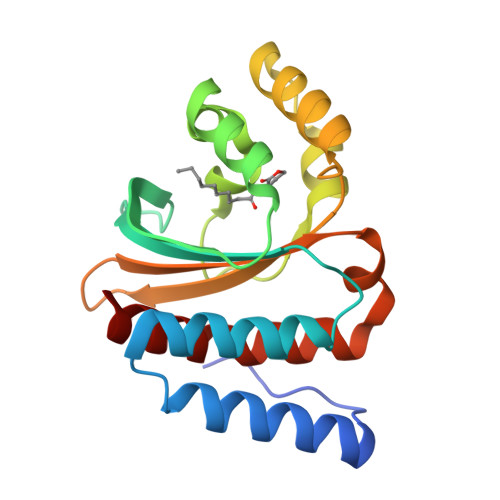
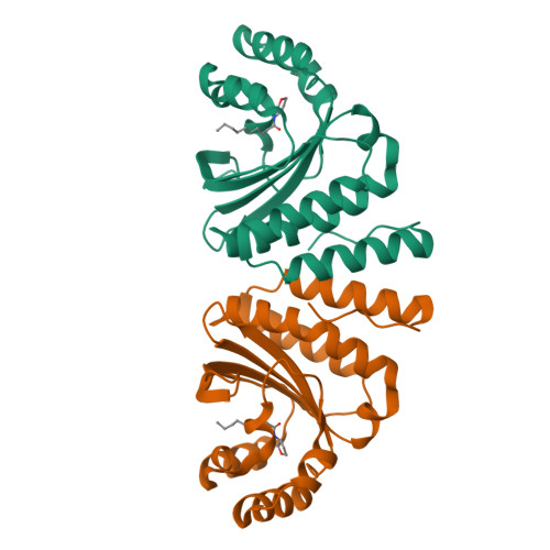
Crystal structure of CviR ligand-binding domain bound to C10-HSL


Experimental Data Snapshot

  • Method: X-RAY DIFFRACTION
  • Resolution: 1.55 Å
  • R-Value Free: 
    0.238 (Depositor), 0.240 (DCC) 
  • R-Value Work: 
    0.209 (Depositor), 0.210 (DCC) 
  • R-Value Observed: 
    0.210 (Depositor) 

Starting Model: experimental
View more details

wwPDB Validation   3D Report Full Report


Ligand Structure Quality Assessment 

Created with Raphaël 2.3.0Worse 01 BetterLigand structure goodness of fit to experimental dataBest fitted HL0Click on this verticalbar to view details

This is version 1.2 of the entry. See complete history


Literature

A strategy for antagonizing quorum sensing.

Chen, G.Swem, L.R.Swem, D.L.Stauff, D.L.O'Loughlin, C.T.Jeffrey, P.D.Bassler, B.L.Hughson, F.M.

(2011) Mol Cell 42: 199-209

  • DOI: https://doi.org/10.1016/j.molcel.2011.04.003
  • Primary Citation of Related Structures:  
    3QP1, 3QP2, 3QP4, 3QP5, 3QP6, 3QP8

  • PubMed Abstract: 

    Quorum-sensing bacteria communicate via small molecules called autoinducers to coordinate collective behaviors. Because quorum sensing controls virulence factor expression in many clinically relevant pathogens, membrane-permeable quorum sensing antagonists that prevent population-wide expression of virulence genes offer a potential route to novel antibacterial therapeutics. Here, we report a strategy for inhibiting quorum-sensing receptors of the widespread LuxR family. Structure-function studies with natural and synthetic ligands demonstrate that the dimeric LuxR-type transcription factor CviR from Chromobacterium violaceum is potently antagonized by molecules that bind in place of the native acylated homoserine lactone autoinducer, provided that they stabilize a closed conformation. In such conformations, each of the two DNA-binding domains interacts with the ligand-binding domain of the opposing monomer. Consequently, the DNA-binding helices are held apart by ∼60 Å, twice the ∼30 Å separation required for operator binding. This approach may represent a general strategy for the inhibition of multidomain proteins.


  • Organizational Affiliation

    Department of Molecular Biology, Princeton University, Princeton, NJ 08544, USA.


Macromolecules
Find similar proteins by:  (by identity cutoff)  |  3D Structure
Entity ID: 1
MoleculeChains Sequence LengthOrganismDetailsImage
CviR transcriptional regulator182Chromobacterium violaceumMutation(s): 0 
Gene Names: cviR
UniProt
Find proteins for D3W065 (Chromobacterium violaceum)
Explore D3W065 
Go to UniProtKB:  D3W065
Entity Groups  
Sequence Clusters30% Identity50% Identity70% Identity90% Identity95% Identity100% Identity
UniProt GroupD3W065
Sequence Annotations
Expand
  • Reference Sequence
Small Molecules
Ligands 1 Unique
IDChains Name / Formula / InChI Key2D Diagram3D Interactions
HL0
Query on HL0

Download Ideal Coordinates CCD File 
B [auth A]N-[(3S)-2-oxotetrahydrofuran-3-yl]decanamide
C14 H25 N O3
TZWZKDULKILUPV-LBPRGKRZSA-N
Experimental Data & Validation

Experimental Data

  • Method: X-RAY DIFFRACTION
  • Resolution: 1.55 Å
  • R-Value Free:  0.238 (Depositor), 0.240 (DCC) 
  • R-Value Work:  0.209 (Depositor), 0.210 (DCC) 
  • R-Value Observed: 0.210 (Depositor) 
Space Group: I 2 2 2
Unit Cell:
Length ( Å )Angle ( ˚ )
a = 55.196α = 90
b = 56.216β = 90
c = 124.944γ = 90
Software Package:
Software NamePurpose
HKL-2000data collection
PHASERphasing
REFMACrefinement
DENZOdata reduction
SCALEPACKdata scaling

Structure Validation

View Full Validation Report


Ligand Structure Quality Assessment 

Created with Raphaël 2.3.0Worse 01 BetterLigand structure goodness of fit to experimental dataBest fitted HL0Click on this verticalbar to view details

Entry History 

Deposition Data

Revision History  (Full details and data files)

  • Version 1.0: 2011-03-30
    Type: Initial release
  • Version 1.1: 2011-07-13
    Changes: Version format compliance
  • Version 1.2: 2023-09-13
    Changes: Data collection, Database references, Derived calculations, Refinement description