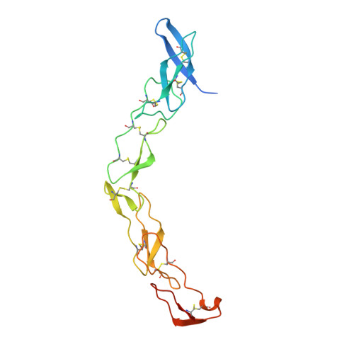
3QO4

The Crystal Structure of Death Receptor 6


Experimental Data Snapshot

  • Method: X-RAY DIFFRACTION
  • Resolution: 2.20 Å
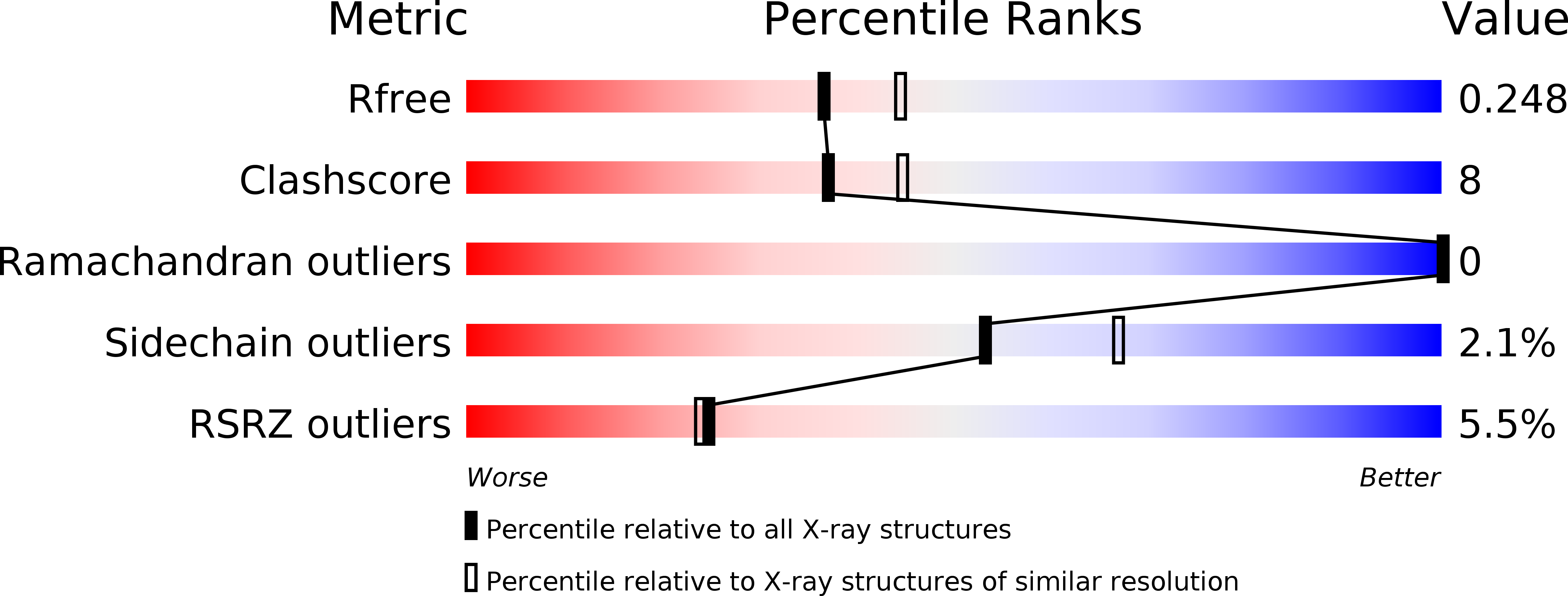
  • R-Value Free: 0.261 
  • R-Value Work: 0.215 

wwPDB Validation   3D Report Full Report


This is version 1.3 of the entry. See complete history


Literature

The crystal structure of death receptor 6 (DR6): a potential receptor of the amyloid precursor protein (APP).

Kuester, M.Kemmerzehl, S.Dahms, S.O.Roeser, D.Than, M.E.

(2011) J Mol Biol 409: 189-201

  • DOI: https://doi.org/10.1016/j.jmb.2011.03.048
  • Primary Citation of Related Structures:  
    3QO4

  • PubMed Abstract: 

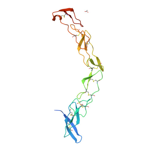
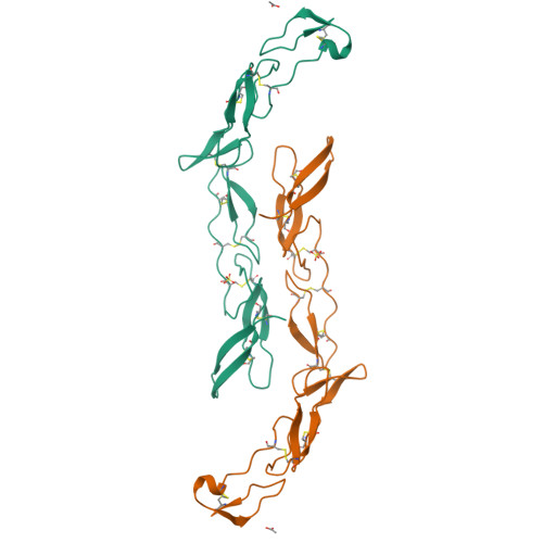
    Death receptors belong to the tumor necrosis factor receptor (TNFR) super family and are intimately involved in the signal transduction during apoptosis, stress response and cellular survival. Here we present the crystal structure of recombinantly expressed death receptor six (DR6), one family member that was recently shown to bind to the amyloid precursor protein (APP) and hence to be probably involved in the development of Alzheimer's disease. The extracellular cysteine rich region of DR6, the typical ligand binding region of all TNFRs, was refined to 2.2 Å resolution and shows that its four constituting cysteine rich domains (CRDs) are arranged in a rod-like overall structure, which presents DR6-specific surface patches responsible for the exclusive recognition of its ligand(s). Based on the structural data, the general ligand binding modes of TNFRs and molecular modeling experiments we were able to elucidate structural features of the potential DR6-APP signaling complex.


  • Organizational Affiliation

    Leibniz Institute for Age Research-Fritz Lipmann Institut, Protein Crystallography Group, Beutenbergstr. 11, 07745 Jena, Germany.


Macromolecules
Find similar proteins by:  (by identity cutoff)  |  3D Structure
Entity ID: 1
MoleculeChains Sequence LengthOrganismDetailsImage
Tumor necrosis factor receptor superfamily member 21181Homo sapiensMutation(s): 0 
Gene Names: TNFRSF21DR6UNQ437/PRO868
UniProt & NIH Common Fund Data Resources
Find proteins for O75509 (Homo sapiens)
Explore O75509 
Go to UniProtKB:  O75509
PHAROS:  O75509
GTEx:  ENSG00000146072 
Entity Groups  
Sequence Clusters30% Identity50% Identity70% Identity90% Identity95% Identity100% Identity
UniProt GroupO75509
Sequence Annotations
Expand
  • Reference Sequence
Small Molecules
Ligands 2 Unique
IDChains Name / Formula / InChI Key2D Diagram3D Interactions
SO4
Query on SO4

Download Ideal Coordinates CCD File 
C [auth A]SULFATE ION
O4 S
QAOWNCQODCNURD-UHFFFAOYSA-L
ACT
Query on ACT

Download Ideal Coordinates CCD File 
B [auth A]ACETATE ION
C2 H3 O2
QTBSBXVTEAMEQO-UHFFFAOYSA-M
Experimental Data & Validation

Experimental Data

  • Method: X-RAY DIFFRACTION
  • Resolution: 2.20 Å
  • R-Value Free: 0.261 
  • R-Value Work: 0.215 
  • Space Group: P 61 2 2
Unit Cell:
Length ( Å )Angle ( ˚ )
a = 77.91α = 90
b = 77.91β = 90
c = 187.844γ = 120
Software Package:
Software NamePurpose
SHARPphasing
CNSrefinement
MOSFLMdata reduction
SCALAdata scaling

Structure Validation

View Full Validation Report


Entry History 

Deposition Data

Revision History  (Full details and data files)

  • Version 1.0: 2011-05-18
    Type: Initial release
  • Version 1.1: 2011-07-13
    Changes: Version format compliance
  • Version 1.2: 2012-02-29
    Changes: Database references
  • Version 1.3: 2024-10-16
    Changes: Data collection, Database references, Derived calculations, Structure summary