3QNG | pdb_00003qng

Crystal Structure Analysis of Lysozyme-bound fac-[Re(CO)3(L-serine)]


Experimental Data Snapshot

  • Method: X-RAY DIFFRACTION
  • Resolution: 1.55 Å
  • R-Value Free: 
    0.217 (Depositor), 0.211 (DCC) 
  • R-Value Work: 
    0.183 (DCC) 
  • R-Value Observed: 
    0.185 (Depositor) 

Starting Model: experimental
View more details

wwPDB Validation 3D Report Full Report

Validation slider image for 3QNG

Ligand Structure Quality Assessment 


This is version 1.3 of the entry. See complete history

Literature

Post-protein-binding reactivity and modifications of the fac-[Re(CO)3]+ core

Zobi, F.Spingler, B.

(2012) Inorg Chem 51: 1210-1212

  • DOI: https://doi.org/10.1021/ic2023314
  • Primary Citation Related Structures: 
    3QE8, 3QNG

  • PubMed Abstract: 

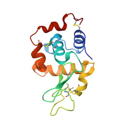
    The reactivity of the [Re(CO)(3)(H(2)O)(2)](+) complex coordinated to the His15 residue of HEW lysozyme is described. In the fully metalated protein (Lys-1), the Re ion retains its reactivity only toward selected ligands, while others induce a ligand-mediated demetalation of the enzyme. It is further shown that some of the complexes that may be "engineered" on the lysozyme do not react with the free protein even if present in solution in excess. The formation of stable metal adducts starting from Lys-1 was confirmed by X-ray crystallography.


  • Organizational Affiliation
    • Institute of Inorganic Chemistry, University of Zürich, Winterthurerstrasse 190, CH-8057 Zürich, Switzerland. fzobi@aci.uzh.ch

Macromolecule Content 

  • Total Structure Weight: 14.83 kDa 
  • Atom Count: 1,120 
  • Modeled Residue Count: 129 
  • Deposited Residue Count: 129 
  • Unique protein chains: 1

Macromolecules

Find similar proteins by:|  3D Structure
Entity ID: 1
MoleculeChains  Sequence LengthOrganismDetailsImage
Lysozyme C129Gallus gallusMutation(s): 0 
EC: 3.2.1.17
UniProt
Find proteins for P00698 (Gallus gallus)
Explore P00698 
Go to UniProtKB:  P00698
Entity Groups
Sequence Clusters30% Identity50% Identity70% Identity90% Identity95% Identity100% Identity
UniProt GroupP00698
Sequence Annotations
Expand
Reference Sequence

Experimental Data & Validation

Experimental Data

  • Method: X-RAY DIFFRACTION
  • Resolution: 1.55 Å
  • R-Value Free:  0.217 (Depositor), 0.211 (DCC) 
  • R-Value Work:  0.183 (DCC) 
  • R-Value Observed: 0.185 (Depositor) 
Space Group: P 43 21 2
Unit Cell:
Length ( Å )Angle ( ˚ )
a = 78.343α = 90
b = 78.343β = 90
c = 36.546γ = 90
Software Package:
Software NamePurpose
RemDAqdata collection
PHASERphasing
SHELXL-97refinement
XDSdata reduction
XDSdata scaling

Structure Validation

View Full Validation Report


Ligand Structure Quality Assessment 


Entry History 

Deposition Data

Revision History  (Full details and data files)

  • Version 1.0: 2012-01-18
    Type: Initial release
  • Version 1.1: 2012-03-21
    Changes: Database references
  • Version 1.2: 2023-11-01
    Changes: Data collection, Database references, Derived calculations, Refinement description
  • Version 1.3: 2024-11-20
    Changes: Structure summary