3QIY

Crystal Structure of BoNT/A LC complexed with Hydroxamate-based Inhibitor PT-1


Experimental Data Snapshot

  • Method: X-RAY DIFFRACTION
  • Resolution: 2.30 Å
  • R-Value Free: 0.263 
  • R-Value Work: 0.226 
  • R-Value Observed: 0.228 

Starting Model: experimental
View more details

wwPDB Validation   3D Report Full Report


Ligand Structure Quality Assessment 


This is version 1.3 of the entry. See complete history


Literature

Structural Characterization of Three Novel Hydroxamate-Based Zinc Chelating Inhibitors of the Clostridium botulinum Serotype A Neurotoxin Light Chain Metalloprotease Reveals a Compact Binding Site Resulting from 60/70 Loop Flexibility.

Thompson, A.A.Jiao, G.S.Kim, S.Thai, A.Cregar-Hernandez, L.Margosiak, S.A.Johnson, A.T.Han, G.W.O'Malley, S.Stevens, R.C.

(2011) Biochemistry 50: 4019-4028

  • DOI: https://doi.org/10.1021/bi2001483
  • Primary Citation of Related Structures:  
    3QIX, 3QIY, 3QIZ, 3QJ0

  • PubMed Abstract: 

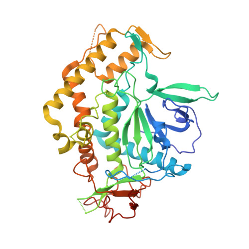
    Neurotoxins synthesized by Clostridium botulinum bacteria (BoNT), the etiological agent of human botulism, are extremely toxic proteins making them high-risk agents for bioterrorism. Small molecule inhibitor development has been focused on the light chain zinc-dependent metalloprotease domain of the neurotoxin, an effort that has been hampered by its relatively flexible active site. Developed in concert with structure--activity relationship studies, the X-ray crystal structures of the complex of BoNT serotype A light chain (BoNT/A LC) with three different micromolar-potency hydroxamate-based inhibitors are reported here. Comparison with an unliganded BoNT/A LC structure reveals significant changes in the active site as a result of binding by the unique inhibitor scaffolds. The 60/70 loop at the opening of the active site pocket undergoes the largest conformational change, presumably through an induced-fit mechanism, resulting in the most compact catalytic pocket observed in all known BoNT/A LC structures.


  • Organizational Affiliation

    Department of Molecular Biology, The Scripps Research Institute, La Jolla, California 92037, United States.


Macromolecules
Find similar proteins by:  (by identity cutoff)  |  3D Structure
Entity ID: 1
MoleculeChains Sequence LengthOrganismDetailsImage
Botulinum neurotoxin type A430Clostridium botulinum A str. HallMutation(s): 0 
Gene Names: botACBO0806CLC_0862Neurotoxin Light Chain
EC: 3.4.24.69
Entity Groups  
Sequence Clusters30% Identity50% Identity70% Identity90% Identity95% Identity100% Identity
Sequence Annotations
Expand
  • Reference Sequence
Small Molecules
Binding Affinity Annotations 
IDSourceBinding Affinity
QI1 PDBBind:  3QIY Ki: 4600 (nM) from 1 assay(s)
Experimental Data & Validation

Experimental Data

  • Method: X-RAY DIFFRACTION
  • Resolution: 2.30 Å
  • R-Value Free: 0.263 
  • R-Value Work: 0.226 
  • R-Value Observed: 0.228 
  • Space Group: P 21 21 2
Unit Cell:
Length ( Å )Angle ( ˚ )
a = 59.075α = 90
b = 190.594β = 90
c = 42.721γ = 90
Software Package:
Software NamePurpose
DENZOdata reduction
SCALEPACKdata scaling
PHASERphasing
BUSTER-TNTrefinement
PDB_EXTRACTdata extraction
Blu-Icedata collection
HKL-2000data reduction
HKL-2000data scaling
BUSTERrefinement

Structure Validation

View Full Validation Report


Ligand Structure Quality Assessment 


Entry History 

Deposition Data

Revision History  (Full details and data files)

  • Version 1.0: 2011-04-13
    Type: Initial release
  • Version 1.1: 2011-07-13
    Changes: Version format compliance
  • Version 1.2: 2017-11-08
    Changes: Refinement description
  • Version 1.3: 2023-09-13
    Changes: Data collection, Database references, Derived calculations, Refinement description