3QIC | pdb_00003qic

The structure of human glucokinase E339K mutation


Experimental Data Snapshot

  • Method: X-RAY DIFFRACTION
  • Resolution: 2.20 Å
  • R-Value Free: 
    0.250 (Depositor), 0.250 (DCC) 
  • R-Value Work: 
    0.213 (Depositor), 0.211 (DCC) 
  • R-Value Observed: 
    0.214 (Depositor) 

Starting Model: experimental
View more details

wwPDB Validation 3D Report Full Report

Validation slider image for 3QIC

Ligand Structure Quality Assessment 


This is version 1.5 of the entry. See complete history

Literature

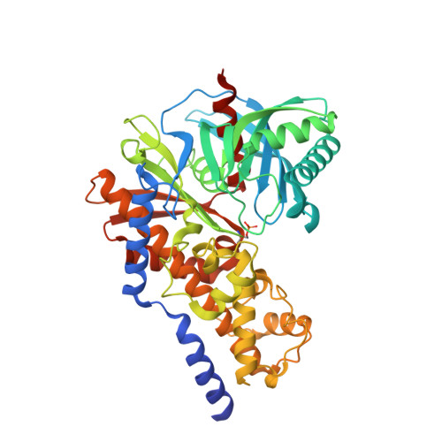
Crystal structure of E339K mutated human glucokinase reveals changes in the ATP binding site.

Liu, Q.Shen, Y.Liu, S.Weng, J.Liu, J.

(2011) FEBS Lett 585: 1175-1179

  • DOI: https://doi.org/10.1016/j.febslet.2011.03.026
  • Primary Citation Related Structures: 
    3QIC

  • PubMed Abstract: 

    Human glucokinase (GK) plays an important role in glucose homeostasis. An E339K mutation in GK was recently found to be associated with hyperglycemia. It showed lower enzyme activity and impaired protein stability compared to the wild-type enzyme. Here, we present the crystal structure of E339K GK in complex with glucose. This mutation results in a conformational change of His416, spatially interfering with adenosine-triphosphate (ATP) binding. Furthermore, Ser411 at the ATP binding site is phosphorylated and then hydrogen bonded with Thr82, physically blocking the ATP binding. These findings provide structural basis for the reduced activity of this mutant.


  • Organizational Affiliation
    • State Key Laboratory of Respiratory Diseases, Guangzhou Institutes of Biomedicine and Health, Chinese Academy of Sciences, Guangzhou 510530, China.

Macromolecule Content 

  • Total Structure Weight: 53.81 kDa 
  • Atom Count: 3,662 
  • Modeled Residue Count: 453 
  • Deposited Residue Count: 470 
  • Unique protein chains: 1

Macromolecules

Find similar proteins by:|  3D Structure
Entity ID: 1
MoleculeChains  Sequence LengthOrganismDetailsImage
Glucokinase470Homo sapiensMutation(s): 1 
Gene Names: GCK
EC: 2.7.1.2 (PDB Primary Data), 2.7.1.1 (UniProt)
UniProt & NIH Common Fund Data Resources
Find proteins for P35557 (Homo sapiens)
Explore P35557 
Go to UniProtKB:  P35557
PHAROS:  P35557
GTEx:  ENSG00000106633 
Entity Groups
Sequence Clusters30% Identity50% Identity70% Identity90% Identity95% Identity100% Identity
UniProt GroupP35557
Sequence Annotations
Expand
Reference Sequence

Small Molecules

Modified Residues  1 Unique
IDChains TypeFormula2D DiagramParent
SEP
Query on SEP
A
L-PEPTIDE LINKINGC3 H8 N O6 PSER

Experimental Data & Validation

Experimental Data

  • Method: X-RAY DIFFRACTION
  • Resolution: 2.20 Å
  • R-Value Free:  0.250 (Depositor), 0.250 (DCC) 
  • R-Value Work:  0.213 (Depositor), 0.211 (DCC) 
  • R-Value Observed: 0.214 (Depositor) 
Space Group: P 43 21 2
Unit Cell:
Length ( Å )Angle ( ˚ )
a = 80.74α = 90
b = 80.74β = 90
c = 178.41γ = 90
Software Package:
Software NamePurpose
MAR345data collection
MOLREPphasing
PHENIXrefinement
MOSFLMdata reduction
SCALAdata scaling

Structure Validation

View Full Validation Report


Ligand Structure Quality Assessment 


Entry History 

Deposition Data

Revision History  (Full details and data files)

  • Version 1.0: 2011-06-01
    Type: Initial release
  • Version 1.1: 2011-07-13
    Changes: Version format compliance
  • Version 1.2: 2017-11-08
    Changes: Refinement description
  • Version 1.3: 2020-07-29
    Type: Remediation
    Reason: Carbohydrate remediation
    Changes: Data collection, Database references, Derived calculations, Structure summary
  • Version 1.4: 2023-11-01
    Changes: Data collection, Database references, Refinement description, Structure summary
  • Version 1.5: 2024-10-30
    Changes: Structure summary