


Engineered Bacterial Mimics of Human Drug Metabolizing Enzyme CYP2C9
Rentmeister, A., Brown, T.R., Snow, C.D., Carbone, M.N., Arnold, F.H.(2011) ChemCatChem
Experimental Data Snapshot
Starting Model: experimental
View more details
(2011) ChemCatChem
Entity ID: 1 | |||||
|---|---|---|---|---|---|
| Molecule | Chains | Sequence Length | Organism | Details | Image |
| Evolved Cytochrome P450 variant (22A3) | A [auth B], B [auth A] | 472 | Priestia megaterium | Mutation(s): 0 EC: 1.14.14.1 | 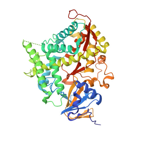 |
| Ligands 1 Unique | |||||
|---|---|---|---|---|---|
| ID | Chains | Name / Formula / InChI Key | 2D Diagram | 3D Interactions | |
| HEM Download:Ideal Coordinates CCD File | C [auth B], D [auth A] | PROTOPORPHYRIN IX CONTAINING FE C34 H32 Fe N4 O4 KABFMIBPWCXCRK-RGGAHWMASA-L |  | ||
| Length ( Å ) | Angle ( ˚ ) |
|---|---|
| a = 82.35 | α = 90 |
| b = 83.77 | β = 90 |
| c = 143.02 | γ = 90 |
| Software Name | Purpose |
|---|---|
| Blu-Ice | data collection |
| PHASER | phasing |
| PHENIX | refinement |
| MOSFLM | data reduction |
| SCALA | data scaling |


