3QGO | pdb_00003qgo

Structure of Thermolysin in complex with L-Phenylalanine methylester


Experimental Data Snapshot

  • Method: X-RAY DIFFRACTION
  • Resolution: 1.45 Å
  • R-Value Free: 
    0.142 (Depositor), 0.141 (DCC) 
  • R-Value Work: 
    0.111 (Depositor), 0.112 (DCC) 
  • R-Value Observed: 
    0.112 (Depositor) 

Starting Model: experimental
View more details

wwPDB Validation 3D Report Full Report

Validation slider image for 3QGO

Ligand Structure Quality Assessment 


This is version 1.4 of the entry. See complete history

Literature

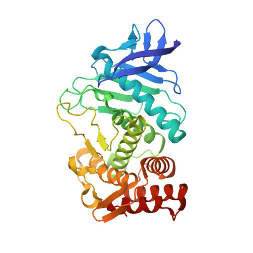
Synthesis of Aspartame by Thermolysin: An X-ray Structural Study.

Birrane, G.Bhyravbhatla, B.Navia, M.A.

(2014) ACS Med Chem Lett 5: 706-710

  • DOI: https://doi.org/10.1021/ml500101z
  • Primary Citation Related Structures: 
    3QGO, 3QH1, 3QH5

  • PubMed Abstract: 

    Protease mediated peptide synthesis (PMPS) was first described in the 1930s but remains underexploited today. In most PMPS, the reaction equilibrium is shifted toward synthesis by the aqueous insolubility of product generated. Substrates and proteases are selected by trial and error, yields are modest, and reaction times are slow. Once implemented, however, PMPS reactions can be simple, environmentally benign, and readily scalable to a commercial level. We examined the PMPS of a precursor of the artificial sweetener aspartame, a multiton peptide synthesis catalyzed by the enzyme thermolysin. X-ray structures of thermolysin in complex with aspartame substrates separately, and after PMPS in a crystal, rationalize the reaction's substrate preferences and reveal an unexpected form of substrate inhibition that explains its sluggishness. Structure guided optimization of this and other PMPS reactions could expand the economic viability of commercial peptides beyond current high-potency, low-volume therapeutics, with substantial green chemistry advantages.


  • Organizational Affiliation
    • Division of Experimental Medicine, Beth Israel Deaconess Medical Center , Boston, Massachusetts 02215, United States.

Macromolecule Content 

  • Total Structure Weight: 35.9 kDa 
  • Atom Count: 3,140 
  • Modeled Residue Count: 316 
  • Deposited Residue Count: 316 
  • Unique protein chains: 1

Macromolecules

Find similar proteins by:|  3D Structure
Entity ID: 1
MoleculeChains  Sequence LengthOrganismDetailsImage
Thermolysin316Bacillus thermoproteolyticusMutation(s): 0 
EC: 3.4.24.27
UniProt
Find proteins for P00800 (Bacillus thermoproteolyticus)
Explore P00800 
Go to UniProtKB:  P00800
Entity Groups
Sequence Clusters30% Identity50% Identity70% Identity90% Identity95% Identity100% Identity
UniProt GroupP00800
Sequence Annotations
Expand
Reference Sequence

Small Molecules

Ligands 6 Unique
IDChains Name / Formula / InChI Key2D Diagram3D Interactions
P6G

Query on P6G


Download:Ideal Coordinates CCD File
M [auth A],
N [auth A]
HEXAETHYLENE GLYCOL
C12 H26 O7
IIRDTKBZINWQAW-UHFFFAOYSA-N
0A9

Query on 0A9


Download:Ideal Coordinates CCD File
G [auth A],
H [auth A],
I [auth A]
methyl L-phenylalaninate
C10 H13 N O2
VSDUZFOSJDMAFZ-VIFPVBQESA-N
DMS

Query on DMS


Download:Ideal Coordinates CCD File
J [auth A],
K [auth A]
DIMETHYL SULFOXIDE
C2 H6 O S
IAZDPXIOMUYVGZ-UHFFFAOYSA-N
ZN

Query on ZN


Download:Ideal Coordinates CCD File
F [auth A]ZINC ION
Zn
PTFCDOFLOPIGGS-UHFFFAOYSA-N
ACT

Query on ACT


Download:Ideal Coordinates CCD File
L [auth A]ACETATE ION
C2 H3 O2
QTBSBXVTEAMEQO-UHFFFAOYSA-M
CA

Query on CA


Download:Ideal Coordinates CCD File
B [auth A],
C [auth A],
D [auth A],
E [auth A]
CALCIUM ION
Ca
BHPQYMZQTOCNFJ-UHFFFAOYSA-N

Experimental Data & Validation

Experimental Data

  • Method: X-RAY DIFFRACTION
  • Resolution: 1.45 Å
  • R-Value Free:  0.142 (Depositor), 0.141 (DCC) 
  • R-Value Work:  0.111 (Depositor), 0.112 (DCC) 
  • R-Value Observed: 0.112 (Depositor) 
Space Group: P 61 2 2
Unit Cell:
Length ( Å )Angle ( ˚ )
a = 93.066α = 90
b = 93.066β = 90
c = 128.171γ = 120
Software Package:
Software NamePurpose
CBASSdata collection
MOLREPphasing
REFMACrefinement
HKL-2000data reduction
HKL-2000data scaling

Structure Validation

View Full Validation Report


Ligand Structure Quality Assessment 


Entry History 

Deposition Data

Revision History  (Full details and data files)

  • Version 1.0: 2012-01-04
    Type: Initial release
  • Version 1.1: 2014-04-30
    Changes: Database references
  • Version 1.2: 2014-07-02
    Changes: Database references
  • Version 1.3: 2023-09-13
    Changes: Data collection, Database references, Derived calculations, Refinement description
  • Version 1.4: 2023-12-06
    Changes: Data collection