3QGH | pdb_00003qgh

Crystal structure of the hepatitis C virus NS5B RNA-dependent RNA polymerase genotype 1a complex with N-cyclopropyl-6-[(3R)-3-{[4-(trifluoromethoxy)benzyl]carbamoyl}-4-{[4-(trifluoromethoxy)phenyl]sulfonyl}piperazin-1-yl]pyridazine-3-carboxamide


Experimental Data Snapshot

  • Method: X-RAY DIFFRACTION
  • Resolution: 2.14 Å
  • R-Value Free: 
    0.204 (Depositor), 0.201 (DCC) 
  • R-Value Work: 
    0.165 (Depositor), 0.163 (DCC) 
  • R-Value Observed: 
    0.166 (Depositor) 

Starting Model: experimental
View more details

wwPDB Validation 3D Report Full Report

Validation slider image for 3QGH

Ligand Structure Quality Assessment 


This is version 1.2 of the entry. See complete history

Literature

Investigation of the mode of binding of a novel series of N-benzyl-4-heteroaryl-1-(phenylsulfonyl)piperazine-2-carboxamides to the hepatitis C virus polymerase.

Gentles, R.G.Sheriff, S.Beno, B.R.Wan, C.Kish, K.Ding, M.Zheng, X.Chupak, L.Poss, M.A.Witmer, M.R.Morin, P.Wang, Y.K.Rigat, K.Lemm, J.Voss, S.Liu, M.Pelosi, L.Roberts, S.B.Gao, M.Kadow, J.F.

(2011) Bioorg Med Chem Lett 21: 2212-2215

  • DOI: https://doi.org/10.1016/j.bmcl.2011.03.011
  • Primary Citation Related Structures: 
    3QGD, 3QGE, 3QGF, 3QGG, 3QGH, 3QGI

  • PubMed Abstract: 

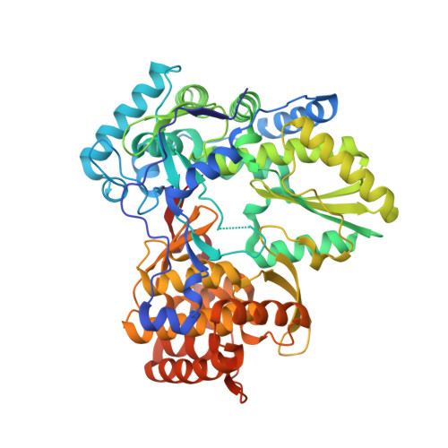
    Structure based rationales for the activities of potent N-benzyl-4-heteroaryl-1-(phenylsulfonyl)piperazine-2-carboxamide inhibitors of the hepatitis C viral polymerase are described herein. These compounds bind to the hepatitis C virus non-structural protein 5B (NS5B), and co-crystal structures of select examples from this series with NS5B are reported. Comparison of co-crystal structures of a potent analog with both NS5B genotype 1a and genotype 1b provides a possible explanation for the genotype-selectivity observed with this compound class and suggests opportunities for the further optimization of the series.


  • Organizational Affiliation
    • Bristol Myers Squibb, Chemical and Protein Technologies, Wallingford, CT 06492, United States. robert.gentles@bms.com

Macromolecule Content 

  • Total Structure Weight: 64.18 kDa 
  • Atom Count: 4,799 
  • Modeled Residue Count: 559 
  • Deposited Residue Count: 571 
  • Unique protein chains: 1

Macromolecules

Find similar proteins by:|  3D Structure
Entity ID: 1
MoleculeChains  Sequence LengthOrganismDetailsImage
RNA-directed RNA polymerase571hepatitis C virus genotype 1aMutation(s): 0 
Gene Names: NS5B
EC: 2.7.7.48
UniProt
Find proteins for P26664 (Hepatitis C virus genotype 1a (isolate 1))
Explore P26664 
Go to UniProtKB:  P26664
Entity Groups
Sequence Clusters30% Identity50% Identity70% Identity90% Identity95% Identity100% Identity
UniProt GroupP26664
Sequence Annotations
Expand
Reference Sequence

Experimental Data & Validation

Experimental Data

  • Method: X-RAY DIFFRACTION
  • Resolution: 2.14 Å
  • R-Value Free:  0.204 (Depositor), 0.201 (DCC) 
  • R-Value Work:  0.165 (Depositor), 0.163 (DCC) 
  • R-Value Observed: 0.166 (Depositor) 
Space Group: P 21 21 21
Unit Cell:
Length ( Å )Angle ( ˚ )
a = 61.3α = 90
b = 84.7β = 90
c = 132.9γ = 90
Software Package:
Software NamePurpose
BUSTER-TNTrefinement
PDB_EXTRACTdata extraction
HKL-2000data reduction
HKL-2000data scaling
AMoREphasing
BUSTERrefinement

Structure Validation

View Full Validation Report


Ligand Structure Quality Assessment 


Entry History 

Deposition Data

  • Released Date: 2011-04-20 
  • Deposition Author(s): Sheriff, S.

Revision History  (Full details and data files)

  • Version 1.0: 2011-04-20
    Type: Initial release
  • Version 1.1: 2011-07-13
    Changes: Version format compliance
  • Version 1.2: 2023-09-13
    Changes: Data collection, Database references, Derived calculations, Refinement description