3QC9 | pdb_00003qc9

Crystal structure of cross-linked bovine GRK1 T8C/N480C double mutant complexed with ADP and Mg


Experimental Data Snapshot

  • Method: X-RAY DIFFRACTION
  • Resolution: 2.70 Å
  • R-Value Free: 
    0.229 (Depositor), 0.225 (DCC) 
  • R-Value Work: 
    0.192 (Depositor), 0.190 (DCC) 
  • R-Value Observed: 
    0.194 (Depositor) 

Starting Model: experimental
View more details

wwPDB Validation 3D Report Full Report

Validation slider image for 3QC9

Ligand Structure Quality Assessment 


This is version 1.2 of the entry. See complete history

Literature

Activation of G Protein-Coupled Receptor Kinase 1 Involves Interactions between Its N-Terminal Region and Its Kinase Domain.

Huang, C.C.Orban, T.Jastrzebska, B.Palczewski, K.Tesmer, J.J.

(2011) Biochemistry 50: 1940-1949

  • DOI: https://doi.org/10.1021/bi101606e
  • Primary Citation Related Structures: 
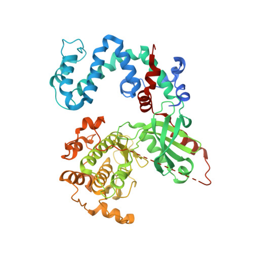
    3QC9

  • PubMed Abstract: 

    G protein-coupled receptor kinases (GRKs) phosphorylate activated G protein-coupled receptors (GPCRs) to initiate receptor desensitization. In addition to the canonical phosphoacceptor site of the kinase domain, activated receptors bind to a distinct docking site that confers higher affinity and activates GRKs allosterically. Recent mutagenesis and structural studies support a model in which receptor docking activates a GRK by stabilizing the interaction of its ∼20-amino acid N-terminal region with the kinase domain. This interaction in turn stabilizes a closed, more active conformation of the enzyme. To investigate the importance of this interaction for the process of GRK activation, we first validated the functionality of the N-terminal region in rhodopsin kinase (GRK1) by site-directed mutagenesis and then introduced a disulfide bond to cross-link the N-terminal region of GRK1 with its specific binding site on the kinase domain. Characterization of the kinetic and biophysical properties of the cross-linked protein showed that disulfide bond formation greatly enhances the catalytic efficiency of the peptide phosphorylation, but receptor-dependent phosphorylation, Meta II stabilization, and inhibition of transducin activation were unaffected. These data indicate that the interaction of the N-terminal region with the kinase domain is important for GRK activation but does not dictate the affinity of GRKs for activated receptors.


  • Organizational Affiliation
    • Life Sciences Institute, University of Michigan, Ann Arbor, Michigan 48109-2216, United States.

Macromolecule Content 

  • Total Structure Weight: 247.77 kDa 
  • Atom Count: 15,806 
  • Modeled Residue Count: 1,935 
  • Deposited Residue Count: 2,172 
  • Unique protein chains: 1

Macromolecules

Find similar proteins by:|  3D Structure
Entity ID: 1
MoleculeChains  Sequence LengthOrganismDetailsImage
Rhodopsin kinase
A, B, C, D
543Bos taurusMutation(s): 2 
Gene Names: GRK1RHOK
EC: 2.7.11.14
UniProt
Find proteins for P28327 (Bos taurus)
Explore P28327 
Go to UniProtKB:  P28327
Entity Groups
Sequence Clusters30% Identity50% Identity70% Identity90% Identity95% Identity100% Identity
UniProt GroupP28327
Sequence Annotations
Expand
Reference Sequence

Small Molecules

Ligands 3 Unique
IDChains Name / Formula / InChI Key2D Diagram3D Interactions
ADP

Query on ADP


Download:Ideal Coordinates CCD File
E [auth A],
I [auth B],
M [auth C],
P [auth D]
ADENOSINE-5'-DIPHOSPHATE
C10 H15 N5 O10 P2
XTWYTFMLZFPYCI-KQYNXXCUSA-N
GOL

Query on GOL


Download:Ideal Coordinates CCD File
H [auth A],
L [auth B]
GLYCEROL
C3 H8 O3
PEDCQBHIVMGVHV-UHFFFAOYSA-N
MG

Query on MG


Download:Ideal Coordinates CCD File
F [auth A]
G [auth A]
J [auth B]
K [auth B]
N [auth C]
F [auth A],
G [auth A],
J [auth B],
K [auth B],
N [auth C],
O [auth C],
Q [auth D],
R [auth D]
MAGNESIUM ION
Mg
JLVVSXFLKOJNIY-UHFFFAOYSA-N

Experimental Data & Validation

Experimental Data

  • Method: X-RAY DIFFRACTION
  • Resolution: 2.70 Å
  • R-Value Free:  0.229 (Depositor), 0.225 (DCC) 
  • R-Value Work:  0.192 (Depositor), 0.190 (DCC) 
  • R-Value Observed: 0.194 (Depositor) 
Space Group: P 1
Unit Cell:
Length ( Å )Angle ( ˚ )
a = 58.696α = 88.07
b = 89.965β = 90.26
c = 122.638γ = 68.78
Software Package:
Software NamePurpose
HKL-2000data collection
PHASERphasing
PHENIXrefinement
HKL-2000data reduction
HKL-2000data scaling

Structure Validation

View Full Validation Report


Ligand Structure Quality Assessment 


Entry History 

Deposition Data

Revision History  (Full details and data files)

  • Version 1.0: 2011-02-16
    Type: Initial release
  • Version 1.1: 2011-07-13
    Changes: Version format compliance
  • Version 1.2: 2023-09-13
    Changes: Data collection, Database references, Derived calculations, Refinement description