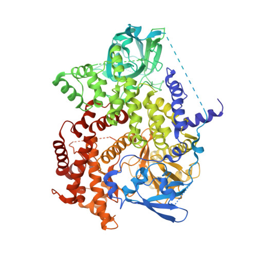
3QAQ

Crystal structure of PI3K-gamma in complex with triazine-benzimidazole 1


Experimental Data Snapshot

  • Method: X-RAY DIFFRACTION
  • Resolution: 2.90 Å
  • R-Value Free: 0.265 
  • R-Value Work: 0.217 
  • R-Value Observed: 0.221 

Starting Model: experimental
View more details

wwPDB Validation   3D Report Full Report


Ligand Structure Quality Assessment 


This is version 1.2 of the entry. See complete history


Literature

Discovery of triazine-benzimidazoles as selective inhibitors of mTOR.

Peterson, E.A.Andrews, P.S.Be, X.Boezio, A.A.Bush, T.L.Cheng, A.C.Coats, J.R.Colletti, A.E.Copeland, K.W.Dupont, M.Graceffa, R.Grubinska, B.Harmange, J.C.Kim, J.L.Mullady, E.L.Olivieri, P.Schenkel, L.B.Stanton, M.K.Teffera, Y.Whittington, D.A.Cai, T.La, D.S.

(2011) Bioorg Med Chem Lett 21: 2064-2070

  • DOI: https://doi.org/10.1016/j.bmcl.2011.02.007
  • Primary Citation of Related Structures:  
    3QAQ, 3QAR

  • PubMed Abstract: 

    mTOR is part of the PI3K/AKT pathway and is a central regulator of cell growth and survival. Since many cancers display mutations linked to the mTOR signaling pathway, mTOR has emerged as an important target for oncology therapy. Herein, we report the discovery of triazine benzimidazole inhibitors that inhibit mTOR kinase activity with up to 200-fold selectivity over the structurally homologous kinase PI3Kα. When tested in a panel of cancer cell lines displaying various mutations, a selective inhibitor from this series inhibited cellular proliferation with a mean IC(50) of 0.41 μM. Lead compound 42 demonstrated up to 83% inhibition of mTOR substrate phosphorylation in a murine pharmacodynamic model.


  • Organizational Affiliation

    Medicinal Chemistry, Amgen Inc, 360 Binney St, Cambridge, MA 02142, USA. epeterso@amgen.com


Macromolecules
Find similar proteins by:  (by identity cutoff)  |  3D Structure
Entity ID: 1
MoleculeChains Sequence LengthOrganismDetailsImage
Phosphatidylinositol-4,5-bisphosphate 3-kinase catalytic subunit gamma isoform960Homo sapiensMutation(s): 0 
Gene Names: PIK3CG
EC: 2.7.1.153 (PDB Primary Data), 2.7.1.137 (UniProt), 2.7.1.154 (UniProt), 2.7.11.1 (UniProt)
UniProt & NIH Common Fund Data Resources
Find proteins for P48736 (Homo sapiens)
Explore P48736 
Go to UniProtKB:  P48736
PHAROS:  P48736
GTEx:  ENSG00000105851 
Entity Groups  
Sequence Clusters30% Identity50% Identity70% Identity90% Identity95% Identity100% Identity
UniProt GroupP48736
Sequence Annotations
Expand
  • Reference Sequence
Small Molecules
Binding Affinity Annotations 
IDSourceBinding Affinity
QAQ BindingDB:  3QAQ Ki: 120 (nM) from 1 assay(s)
IC50: 100 (nM) from 1 assay(s)
PDBBind:  3QAQ IC50: 100 (nM) from 1 assay(s)
Experimental Data & Validation

Experimental Data

  • Method: X-RAY DIFFRACTION
  • Resolution: 2.90 Å
  • R-Value Free: 0.265 
  • R-Value Work: 0.217 
  • R-Value Observed: 0.221 
  • Space Group: C 1 2 1
Unit Cell:
Length ( Å )Angle ( ˚ )
a = 145.395α = 90
b = 68.579β = 94.69
c = 106.501γ = 90
Software Package:
Software NamePurpose
SCALEPACKdata scaling
REFMACrefinement
PDB_EXTRACTdata extraction
ADSCdata collection
HKL-2000data reduction
EPMRphasing

Structure Validation

View Full Validation Report


Ligand Structure Quality Assessment 


Entry History 

Deposition Data

Revision History  (Full details and data files)

  • Version 1.0: 2011-03-30
    Type: Initial release
  • Version 1.1: 2011-07-13
    Changes: Version format compliance
  • Version 1.2: 2023-09-13
    Changes: Data collection, Database references, Derived calculations, Refinement description