3QAL

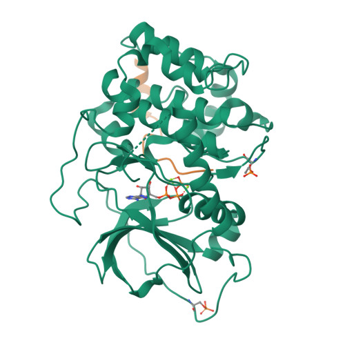
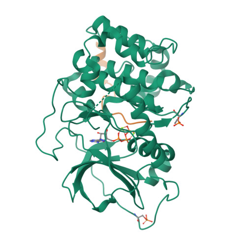
Crystal Structure of Arg280Ala mutant of Catalytic subunit of cAMP-dependent Protein Kinase


Experimental Data Snapshot

  • Method: X-RAY DIFFRACTION
  • Resolution: 1.70 Å
  • R-Value Free: 0.194 
  • R-Value Work: 0.176 

Starting Model: experimental
View more details

wwPDB Validation   3D Report Full Report


Ligand Structure Quality Assessment 


This is version 1.3 of the entry. See complete history


Literature

A conserved Glu-Arg salt bridge connects coevolved motifs that define the eukaryotic protein kinase fold.

Yang, J.Wu, J.Steichen, J.M.Kornev, A.P.Deal, M.S.Li, S.Sankaran, B.Woods, V.L.Taylor, S.S.

(2012) J Mol Biol 415: 666-679

  • DOI: https://doi.org/10.1016/j.jmb.2011.11.035
  • Primary Citation of Related Structures:  
    3QAL, 3QAM

  • PubMed Abstract: 

    Eukaryotic protein kinases (EPKs) feature two coevolved structural segments, the Activation segment, which starts with the Asp-Phe-Gly (DFG) and ends with the Ala-Pro-Glu (APE) motifs, and the helical GHI subdomain that comprises αG-αH-αI helices. Eukaryotic-like kinases have a much shorter Activation segment and lack the GHI subdomain. They thus lack the conserved salt bridge interaction between the APE Glu and an Arg from the GHI subdomain, a hallmark signature of EPKs. Although the conservation of this salt bridge in EPKs is well known and its implication in diseases has been illustrated by polymorphism analysis, its function has not been carefully studied. In this work, we use murine cAMP-dependent protein kinase (protein kinase A) as the model enzyme (Glu208 and Arg280) to examine the role of these two residues. We showed that Ala replacement of either residue caused a 40- to 120-fold decrease in catalytic efficiency of the enzyme due to an increase in K(m)(ATP) and a decrease in k(cat). Crystal structures, as well as solution studies, also demonstrate that this ion pair contributes to the hydrophobic network and stability of the enzyme. We show that mutation of either Glu or Arg to Ala renders both mutant proteins less effective substrates for upstream kinase phosphoinositide-dependent kinase 1. We propose that the Glu208-Arg280 pair serves as a center hub of connectivity between these two structurally conserved elements in EPKs. Mutations of either residue disrupt communication not only between the two segments but also within the rest of the molecule, leading to altered catalytic activity and enzyme regulation.


  • Organizational Affiliation

    Department of Chemistry and Biochemistry, University of California San Diego, San Diego, CA 92093, USA.


Macromolecules
Find similar proteins by:  (by identity cutoff)  |  3D Structure
Entity ID: 1
MoleculeChains Sequence LengthOrganismDetailsImage
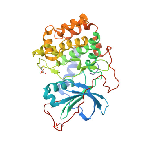
cAMP-dependent protein kinase catalytic subunit alphaA [auth E]350Mus musculusMutation(s): 1 
Gene Names: PrkacaPkaca
EC: 2.7.11.11
UniProt
Find proteins for P05132 (Mus musculus)
Explore P05132 
Go to UniProtKB:  P05132
Entity Groups  
Sequence Clusters30% Identity50% Identity70% Identity90% Identity95% Identity100% Identity
UniProt GroupP05132
Sequence Annotations
Expand
  • Reference Sequence

Find similar proteins by:  Sequence   |   3D Structure  

Entity ID: 2
MoleculeChains Sequence LengthOrganismDetailsImage
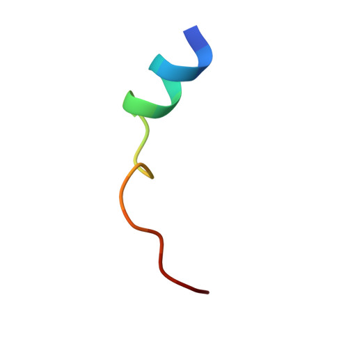
Protein kinase inhibitorB [auth I]18Mus musculusMutation(s): 0 
Gene Names: Pkia
UniProt & NIH Common Fund Data Resources
Find proteins for P63248 (Mus musculus)
Explore P63248 
Go to UniProtKB:  P63248
IMPC:  MGI:104747
Entity Groups  
Sequence Clusters30% Identity50% Identity70% Identity90% Identity95% Identity100% Identity
UniProt GroupP63248
Sequence Annotations
Expand
  • Reference Sequence
Small Molecules
Modified Residues  2 Unique
IDChains TypeFormula2D DiagramParent
SEP
Query on SEP
A [auth E]L-PEPTIDE LINKINGC3 H8 N O6 PSER
TPO
Query on TPO
A [auth E]L-PEPTIDE LINKINGC4 H10 N O6 PTHR
Experimental Data & Validation

Experimental Data

  • Method: X-RAY DIFFRACTION
  • Resolution: 1.70 Å
  • R-Value Free: 0.194 
  • R-Value Work: 0.176 
  • Space Group: P 21 21 21
Unit Cell:
Length ( Å )Angle ( ˚ )
a = 57.758α = 90
b = 80.082β = 90
c = 97.715γ = 90
Software Package:
Software NamePurpose
ADSCdata collection
AMoREphasing
CNSrefinement
HKL-2000data reduction
SCALEPACKdata scaling

Structure Validation

View Full Validation Report


Ligand Structure Quality Assessment 


Entry History 

Deposition Data

Revision History  (Full details and data files)

  • Version 1.0: 2011-12-07
    Type: Initial release
  • Version 1.1: 2013-06-19
    Changes: Database references
  • Version 1.2: 2023-09-13
    Changes: Data collection, Database references, Derived calculations, Refinement description
  • Version 1.3: 2024-10-30
    Changes: Structure summary