3Q9F | pdb_00003q9f

Crystal Structure of APAH complexed with CAPS


Experimental Data Snapshot

  • Method: X-RAY DIFFRACTION
  • Resolution: 2.35 Å
  • R-Value Free: 
    0.225 (Depositor), 0.225 (DCC) 
  • R-Value Work: 
    0.189 (Depositor), 0.189 (DCC) 
  • R-Value Observed: 
    0.189 (Depositor) 

Starting Model: experimental
View more details

wwPDB Validation 3D Report Full Report

Validation slider image for 3Q9F

Ligand Structure Quality Assessment 


This is version 1.3 of the entry. See complete history

Literature

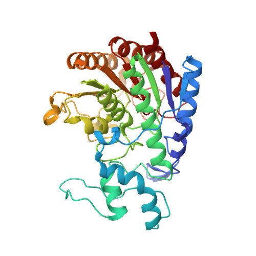
Structure of prokaryotic polyamine deacetylase reveals evolutionary functional relationships with eukaryotic histone deacetylases .

Lombardi, P.M.Angell, H.D.Whittington, D.A.Flynn, E.F.Rajashankar, K.R.Christianson, D.W.

(2011) Biochemistry 50: 1808-1817

  • DOI: https://doi.org/10.1021/bi101859k
  • Primary Citation Related Structures: 
    3Q9B, 3Q9C, 3Q9E, 3Q9F

  • PubMed Abstract: 

    Polyamines are a ubiquitous class of polycationic small molecules that can influence gene expression by binding to nucleic acids. Reversible polyamine acetylation regulates nucleic acid binding and is required for normal cell cycle progression and proliferation. Here, we report the structures of Mycoplana ramosa acetylpolyamine amidohydrolase (APAH) complexed with a transition state analogue and a hydroxamate inhibitor and an inactive mutant complexed with two acetylpolyamine substrates. The structure of APAH is the first of a histone deacetylase-like oligomer and reveals that an 18-residue insert in the L2 loop promotes dimerization and the formation of an 18 Å long "L"-shaped active site tunnel at the dimer interface, accessible only to narrow and flexible substrates. The importance of dimerization for polyamine deacetylase function leads to the suggestion that a comparable dimeric or double-domain histone deacetylase could catalyze polyamine deacetylation reactions in eukaryotes.


  • Organizational Affiliation
    • Roy and Diana Vagelos Laboratories, Department of Chemistry, University of Pennsylvania, 231 South 34th Street, Philadelphia, Pennsylvania 19104-6323, United States.

Macromolecule Content 

  • Total Structure Weight: 442.08 kDa 
  • Atom Count: 32,471 
  • Modeled Residue Count: 4,090 
  • Deposited Residue Count: 4,092 
  • Unique protein chains: 1

Macromolecules

Find similar proteins by:|  3D Structure
Entity ID: 1
MoleculeChains  Sequence LengthOrganismDetailsImage
Acetylpolyamine amidohydrolase
A, B, C, D, E
A, B, C, D, E, F, G, H, I, J, K, L
341Mycoplana ramosaMutation(s): 0 
Gene Names: aphAaph
EC: 3.5.1.62 (UniProt), 3.5.1 (UniProt), 3.5.1.48 (UniProt)
UniProt
Find proteins for Q48935 (Mycoplana ramosa)
Explore Q48935 
Go to UniProtKB:  Q48935
Entity Groups
Sequence Clusters30% Identity50% Identity70% Identity90% Identity95% Identity100% Identity
UniProt GroupQ48935
Sequence Annotations
Expand
Reference Sequence

Small Molecules

Ligands 5 Unique
IDChains Name / Formula / InChI Key2D Diagram3D Interactions
CXS

Query on CXS


Download:Ideal Coordinates CCD File
BB [auth H]
DA [auth D]
HB [auth I]
JA [auth E]
M [auth A]
BB [auth H],
DA [auth D],
HB [auth I],
JA [auth E],
M [auth A],
MB [auth J],
PA [auth F],
RB [auth K],
S [auth B],
VA [auth G],
WB [auth L],
X [auth C]
3-CYCLOHEXYL-1-PROPYLSULFONIC ACID
C9 H19 N O3 S
PJWWRFATQTVXHA-UHFFFAOYSA-N
PO4

Query on PO4


Download:Ideal Coordinates CCD File
CB [auth H]
EA [auth D]
IB [auth I]
KA [auth E]
N [auth A]
CB [auth H],
EA [auth D],
IB [auth I],
KA [auth E],
N [auth A],
NB [auth J],
QA [auth F],
SB [auth K],
T [auth B],
WA [auth G],
XB [auth L],
Y [auth C]
PHOSPHATE ION
O4 P
NBIIXXVUZAFLBC-UHFFFAOYSA-K
ZN

Query on ZN


Download:Ideal Coordinates CCD File
AB [auth G]
BC [auth L]
CA [auth C]
GB [auth H]
IA [auth D]
AB [auth G],
BC [auth L],
CA [auth C],
GB [auth H],
IA [auth D],
LB [auth I],
OA [auth E],
QB [auth J],
R [auth A],
UA [auth F],
VB [auth K],
W [auth B]
ZINC ION
Zn
PTFCDOFLOPIGGS-UHFFFAOYSA-N
K

Query on K


Download:Ideal Coordinates CCD File
AC [auth L]
BA [auth C]
DB [auth H]
FA [auth D]
FB [auth H]
AC [auth L],
BA [auth C],
DB [auth H],
FA [auth D],
FB [auth H],
HA [auth D],
JB [auth I],
LA [auth E],
NA [auth E],
O [auth A],
OB [auth J],
Q [auth A],
RA [auth F],
TA [auth F],
TB [auth K],
U [auth B],
XA [auth G],
YB [auth L],
Z [auth C],
ZA [auth G]
POTASSIUM ION
K
NPYPAHLBTDXSSS-UHFFFAOYSA-N
NA

Query on NA


Download:Ideal Coordinates CCD File
AA [auth C]
EB [auth H]
GA [auth D]
KB [auth I]
MA [auth E]
AA [auth C],
EB [auth H],
GA [auth D],
KB [auth I],
MA [auth E],
P [auth A],
PB [auth J],
SA [auth F],
UB [auth K],
V [auth B],
YA [auth G],
ZB [auth L]
SODIUM ION
Na
FKNQFGJONOIPTF-UHFFFAOYSA-N

Experimental Data & Validation

Experimental Data

  • Method: X-RAY DIFFRACTION
  • Resolution: 2.35 Å
  • R-Value Free:  0.225 (Depositor), 0.225 (DCC) 
  • R-Value Work:  0.189 (Depositor), 0.189 (DCC) 
  • R-Value Observed: 0.189 (Depositor) 
Space Group: P 1
Unit Cell:
Length ( Å )Angle ( ˚ )
a = 118.252α = 98.34
b = 119.651β = 94.94
c = 119.571γ = 114.95
Software Package:
Software NamePurpose
CNSrefinement
PDB_EXTRACTdata extraction
APS-24IDCdata collection
HKL-2000data reduction
HKL-2000data scaling
CNSphasing

Structure Validation

View Full Validation Report


Ligand Structure Quality Assessment 


Entry History 

Deposition Data

Revision History  (Full details and data files)

  • Version 1.0: 2011-03-02
    Type: Initial release
  • Version 1.1: 2011-07-13
    Changes: Version format compliance
  • Version 1.2: 2024-02-21
    Changes: Data collection, Database references, Derived calculations
  • Version 1.3: 2024-04-03
    Changes: Refinement description