3Q85 | pdb_00003q85

Crystal Structure of Rem2 G-domain -GTP Analog Complex


Experimental Data Snapshot

  • Method: X-RAY DIFFRACTION
  • Resolution: 1.76 Å
  • R-Value Free: 
    0.235 (Depositor), 0.231 (DCC) 
  • R-Value Work: 
    0.208 (Depositor), 0.203 (DCC) 
  • R-Value Observed: 
    0.209 (Depositor) 

Starting Model: experimental
View more details

wwPDB Validation 3D Report Full Report

Validation slider image for 3Q85

Ligand Structure Quality Assessment 


This is version 1.2 of the entry. See complete history

Literature

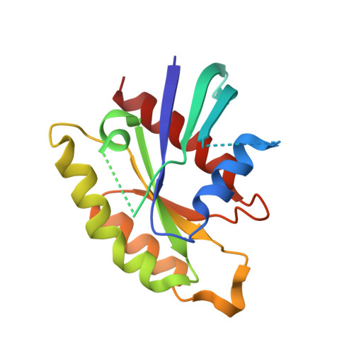
RGK Family G-Domain:GTP Analog Complex Structures and Nucleotide-Binding Properties.

Sasson, Y.Navon-Perry, L.Huppert, D.Hirsch, J.A.

(2011) J Mol Biology 413: 372-389

  • DOI: https://doi.org/10.1016/j.jmb.2011.08.017
  • Primary Citation Related Structures: 
    3Q72, 3Q7P, 3Q7Q, 3Q85

  • PubMed Abstract: 

    The RGK family of small G-proteins, including Rad, Gem, Rem1, and Rem2, is inducibly expressed in various mammalian tissues and interacts with voltage-dependent calcium channels and Rho kinase. Many questions remain regarding their physiological roles and molecular mechanism. Previous crystallographic studies reported RGK G-domain:guanosine di-phosphate structures. To test whether RGK proteins undergo a nucleotide-induced conformational change, we determined the crystallographic structures of Rad:GppNHp and Rem2:GppNHp to 1.7 and 1.8 Å resolutions, respectively. Also, we characterized the nucleotide-binding properties and conformations for Gem, Rad, and several structure-based mutants using fluorescence spectroscopy. The results suggest that RGK G-proteins may not behave as Ras-like canonical nucleotide-induced molecular switches. Further, the RGK proteins have differing structures and nucleotide-binding properties, which may have implications for their varied action on effectors.


  • Organizational Affiliation
    • Department of Biochemistry, Tel Aviv University, Ramat Aviv, Tel Aviv 69978, Israel.

Macromolecule Content 

  • Total Structure Weight: 39.1 kDa 
  • Atom Count: 2,866 
  • Modeled Residue Count: 300 
  • Deposited Residue Count: 338 
  • Unique protein chains: 1

Macromolecules

Find similar proteins by:|  3D Structure
Entity ID: 1
MoleculeChains  Sequence LengthOrganismDetailsImage
GTP-binding protein REM 2
A, B
169Mus musculusMutation(s): 0 
Gene Names: Rem2
UniProt
Find proteins for Q8VEL9 (Mus musculus)
Explore Q8VEL9 
Go to UniProtKB:  Q8VEL9
Entity Groups
Sequence Clusters30% Identity50% Identity70% Identity90% Identity95% Identity100% Identity
UniProt GroupQ8VEL9
Sequence Annotations
Expand
Reference Sequence

Small Molecules

Ligands 3 Unique
IDChains Name / Formula / InChI Key2D Diagram3D Interactions
GNP

Query on GNP


Download:Ideal Coordinates CCD File
C [auth A],
F [auth B]
PHOSPHOAMINOPHOSPHONIC ACID-GUANYLATE ESTER
C10 H17 N6 O13 P3
UQABYHGXWYXDTK-UUOKFMHZSA-N
SCN

Query on SCN


Download:Ideal Coordinates CCD File
E [auth A],
H [auth B],
I [auth B]
THIOCYANATE ION
C N S
ZMZDMBWJUHKJPS-UHFFFAOYSA-M
MG

Query on MG


Download:Ideal Coordinates CCD File
D [auth A],
G [auth B]
MAGNESIUM ION
Mg
JLVVSXFLKOJNIY-UHFFFAOYSA-N

Experimental Data & Validation

Experimental Data

  • Method: X-RAY DIFFRACTION
  • Resolution: 1.76 Å
  • R-Value Free:  0.235 (Depositor), 0.231 (DCC) 
  • R-Value Work:  0.208 (Depositor), 0.203 (DCC) 
  • R-Value Observed: 0.209 (Depositor) 
Space Group: P 1 21 1
Unit Cell:
Length ( Å )Angle ( ˚ )
a = 52.525α = 90
b = 59.08β = 101.26
c = 56.144γ = 90
Software Package:
Software NamePurpose
HKL-2000data collection
PHENIXmodel building
PHENIXrefinement
HKL-2000data reduction
HKL-2000data scaling
PHENIXphasing

Structure Validation

View Full Validation Report


Ligand Structure Quality Assessment 


Entry History 

Deposition Data

Revision History  (Full details and data files)

  • Version 1.0: 2011-09-21
    Type: Initial release
  • Version 1.1: 2011-10-26
    Changes: Database references
  • Version 1.2: 2023-09-13
    Changes: Data collection, Database references, Derived calculations, Refinement description