


3Q58 | pdb_00003q58
3Q58 | pdb_00003q58
Structure of N-acetylmannosamine-6-Phosphate Epimerase from Salmonella enterica
- PDB DOI: https://doi.org/10.2210/pdb3Q58/pdb
- Classification: ISOMERASE
- Organism(s): Salmonella enterica subsp. enterica serovar Typhi
- Expression System: Escherichia coli
- Mutation(s): No
- Deposited: 2010-12-27 Released: 2011-01-12
Experimental Data Snapshot
- Method: X-RAY DIFFRACTION
- Resolution: 1.80 Å
- R-Value Free: 0.204 (Depositor), 0.204 (DCC)
- R-Value Work: 0.174 (Depositor), 0.179 (DCC)
- R-Value Observed: 0.176 (Depositor)
wwPDB Validation 3D Report Full Report

This is version 1.2 of the entry. See complete history.

Macromolecules
Find similar proteins by:
| 3D Structure
Entity ID: 1 | |||||
|---|---|---|---|---|---|
| Molecule | Chains | Sequence Length | Organism | Details | Image |
| N-acetylmannosamine-6-phosphate 2-epimerase | 229 | Salmonella enterica subsp. enterica serovar Typhi | Mutation(s): 0 Gene Names: nanE, STY1166, t1791 EC: 5.1.3.9 | 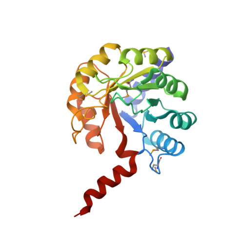 | |
UniProt | |||||
Entity Groups | |||||
| Sequence Clusters | 30% Identity50% Identity70% Identity90% Identity95% Identity100% Identity | ||||
| UniProt Group | P60668 | ||||
Sequence AnnotationsExpand | |||||
Reference Sequence | |||||
Small Molecules
| Ligands 3 Unique | |||||
|---|---|---|---|---|---|
| ID | Chains | Name / Formula / InChI Key | 2D Diagram | 3D Interactions | |
| BTB Download:Ideal Coordinates CCD File | U [auth B] | 2-[BIS-(2-HYDROXY-ETHYL)-AMINO]-2-HYDROXYMETHYL-PROPANE-1,3-DIOL C8 H19 N O5 OWMVSZAMULFTJU-UHFFFAOYSA-N |  | ||
| PEG Download:Ideal Coordinates CCD File | C [auth A] D [auth A] E [auth A] F [auth A] G [auth A] | DI(HYDROXYETHYL)ETHER C4 H10 O3 MTHSVFCYNBDYFN-UHFFFAOYSA-N |  | ||
| CL Download:Ideal Coordinates CCD File | V [auth B] | CHLORIDE ION Cl VEXZGXHMUGYJMC-UHFFFAOYSA-M |  | ||
| Modified Residues 1 Unique | |||||
|---|---|---|---|---|---|
| ID | Chains | Type | Formula | 2D Diagram | Parent |
| MSE Query on MSE | A, B | L-PEPTIDE LINKING | C5 H11 N O2 Se |  | MET |
Experimental Data & Validation
Experimental Data
- Method: X-RAY DIFFRACTION
- Resolution: 1.80 Å
- R-Value Free: 0.204 (Depositor), 0.204 (DCC)
- R-Value Work: 0.174 (Depositor), 0.179 (DCC)
- R-Value Observed: 0.176 (Depositor)
Diffraction Data:
Space Group: P 21 21 2
Unit Cell:
| Length ( Å ) | Angle ( ˚ ) |
|---|---|
| a = 81.153 | α = 90 |
| b = 139.31 | β = 90 |
| c = 38.423 | γ = 90 |
| Software Name | Purpose |
|---|---|
| PHENIX | refinement |
| PDB_EXTRACT | data extraction |
| BLU-MAX | data collection |
| HKL-2000 | data reduction |
| HKL-2000 | data scaling |
| PHASER | phasing |
Entry History
Deposition Data
- Released Date: 2011-01-12 Deposition Author(s): Anderson, S.M., Wawrzak, Z., Kudritska, M., Kwon, K., Anderson, W.F., Savchenko, A., Center for Structural Genomics of Infectious Diseases (CSGID)
Revision History (Full details and data files)
- Version 1.0: 2011-01-12
Type: Initial release - Version 1.1: 2011-07-13
Changes: Version format compliance - Version 1.2: 2025-03-26
Changes: Data collection, Database references, Derived calculations, Structure summary


