3Q4C

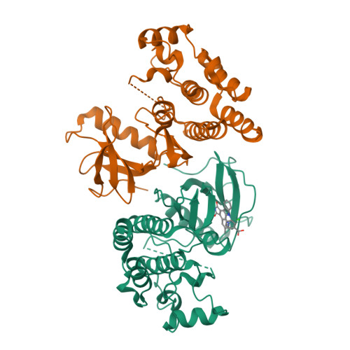
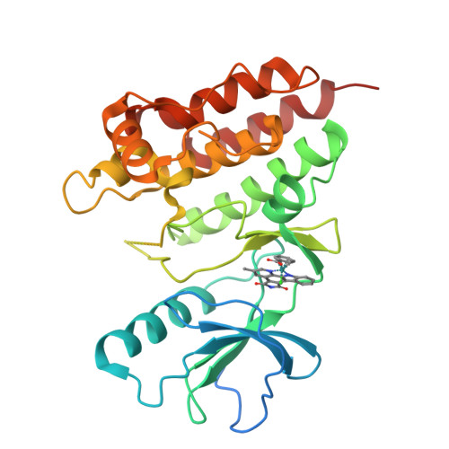
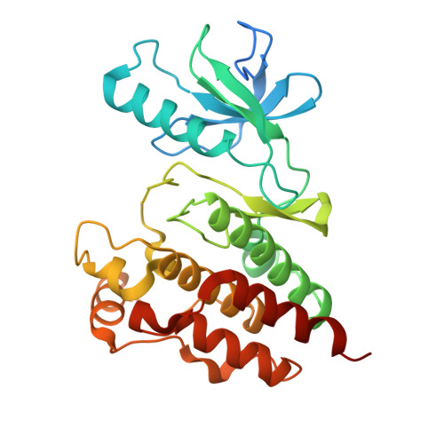
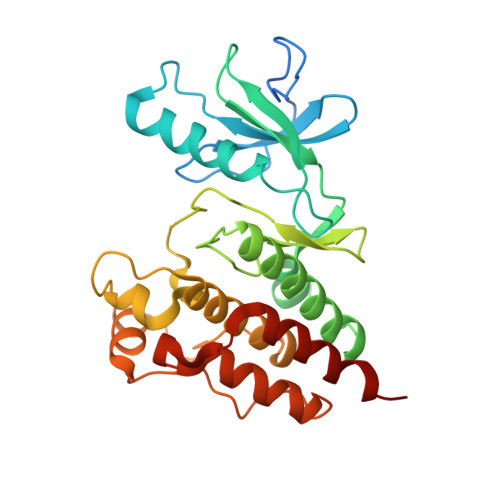
Crystal Structure of Wild Type BRAF kinase domain in complex with organometallic inhibitor CNS292


Experimental Data Snapshot

  • Method: X-RAY DIFFRACTION
  • Resolution: 3.20 Å
  • R-Value Free: 0.280 
  • R-Value Work: 0.232 

wwPDB Validation   3D Report Full Report


Ligand Structure Quality Assessment 


This is version 1.2 of the entry. See complete history


Literature

The crystal structure of BRAF in complex with an organoruthenium inhibitor reveals a mechanism for inhibition of an active form of BRAF kinase.

Xie, P.Streu, C.Qin, J.Bregman, H.Pagano, N.Meggers, E.Marmorstein, R.

(2009) Biochemistry 48: 5187-5198

  • DOI: https://doi.org/10.1021/bi802067u
  • Primary Citation of Related Structures:  
    3Q4C

  • PubMed Abstract: 

    Substitution mutations in the BRAF serine/threonine kinase are found in a variety of human cancers. Such mutations occur in approximately 70% of human malignant melanomas, and a single hyperactivating V600E mutation is found in the activation segment of the kinase domain and accounts for more than 90% of these mutations. Given this correlation, the molecular mechanism for BRAF regulation as well as oncogenic activation has attracted considerable interest, and activated forms of BRAF, such as BRAF(V600E), have become attractive targets for small molecule inhibition. Here we report on the identification and subsequent optimization of a potent BRAF inhibitor, CS292, based on an organometallic kinase inhibitor scaffold. A cocrystal structure of CS292 in complex with the BRAF kinase domain reveals that CS292 binds to the ATP binding pocket of the kinase and is an ATP competitive inhibitor. The structure of the kinase-inhibitor complex also demonstrates that CS292 binds to BRAF in an active conformation and suggests a mechanism for regulation of BRAF by phosphorylation and BRAF(V600E) oncogene-induced activation. The structure of CS292 bound to the active form of the BRAF kinase also provides a novel scaffold for the design of BRAF(V600E) oncogene selective BRAF inhibitors for therapeutic application.


  • Organizational Affiliation

    The Wistar Institute, Philadelphia, Pennsylvania 19104-6323, USA.


Macromolecules
Find similar proteins by:  (by identity cutoff)  |  3D Structure
Entity ID: 1
MoleculeChains Sequence LengthOrganismDetailsImage
Serine/threonine-protein kinase B-raf
A, B
307Homo sapiensMutation(s): 0 
Gene Names: BRAFBRAF1RAFB1
EC: 2.7.11.1
UniProt & NIH Common Fund Data Resources
Find proteins for P15056 (Homo sapiens)
Explore P15056 
Go to UniProtKB:  P15056
PHAROS:  P15056
GTEx:  ENSG00000157764 
Entity Groups  
Sequence Clusters30% Identity50% Identity70% Identity90% Identity95% Identity100% Identity
UniProt GroupP15056
Sequence Annotations
Expand
  • Reference Sequence
Small Molecules
Ligands 1 Unique
IDChains Name / Formula / InChI Key2D Diagram3D Interactions
RSW
Query on RSW

Download Ideal Coordinates CCD File 
C [auth A][(1,2,3,4,5,6-eta)-(1S,2R,3R,4R,5S,6S)-1-carboxycyclohexane-1,2,3,4,5,6-hexayl](chloro)(3-methyl-5,7-dioxo-6,7-dihydro-5H-pyrido[2,3-a]pyrrolo[3,4-c]carbazol-12-ide-kappa~2~N~1~,N~12~)ruthenium(1+)
C25 H16 Cl N3 O4 Ru
LOEHCDRAMDJWEG-UHFFFAOYSA-L
Binding Affinity Annotations 
IDSourceBinding Affinity
RSW PDBBind:  3Q4C IC50: 370 (nM) from 1 assay(s)
Experimental Data & Validation

Experimental Data

  • Method: X-RAY DIFFRACTION
  • Resolution: 3.20 Å
  • R-Value Free: 0.280 
  • R-Value Work: 0.232 
  • Space Group: P 41 21 2
Unit Cell:
Length ( Å )Angle ( ˚ )
a = 94.905α = 90
b = 94.905β = 90
c = 163.349γ = 90
Software Package:
Software NamePurpose
HKL-2000data collection
MOLREPphasing
CNSrefinement
HKL-2000data reduction
HKL-2000data scaling

Structure Validation

View Full Validation Report


Ligand Structure Quality Assessment 


Entry History 

Deposition Data

Revision History  (Full details and data files)

  • Version 1.0: 2011-03-02
    Type: Initial release
  • Version 1.1: 2011-07-13
    Changes: Version format compliance
  • Version 1.2: 2024-02-21
    Changes: Data collection, Database references, Derived calculations