3Q47 | pdb_00003q47

Crystal structure of TPR domain of CHIP complexed with pseudophosphorylated Smad1 peptide


Experimental Data Snapshot

  • Method: X-RAY DIFFRACTION
  • Resolution: 1.71 Å
  • R-Value Free: 
    0.192 (Depositor), 0.191 (DCC) 
  • R-Value Work: 
    0.173 (Depositor), 0.171 (DCC) 
  • R-Value Observed: 
    0.174 (Depositor) 

Starting Model: experimental
View more details

wwPDB Validation 3D Report Full Report

Validation slider image for 3Q47

This is version 1.2 of the entry. See complete history

Literature

Molecular Mechanism of the Negative Regulation of Smad1/5 Protein by Carboxyl Terminus of Hsc70-interacting Protein (CHIP).

Wang, L.Liu, Y.T.Hao, R.Chen, L.Chang, Z.Wang, H.R.Wang, Z.X.Wu, J.W.

(2011) J Biological Chem 286: 15883-15894

  • DOI: https://doi.org/10.1074/jbc.M110.201814
  • Primary Citation Related Structures: 
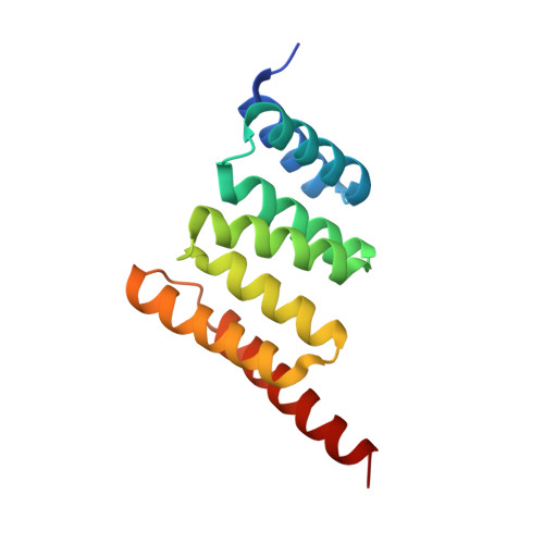
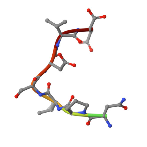
    3Q47, 3Q49, 3Q4A

  • PubMed Abstract: 

    The transforming growth factor-β (TGF-β) superfamily of ligands signals along two intracellular pathways, Smad2/3-mediated TGF-β/activin pathway and Smad1/5/8-mediated bone morphogenetic protein pathway. The C terminus of Hsc70-interacting protein (CHIP) serves as an E3 ubiquitin ligase to mediate the degradation of Smad proteins and many other signaling proteins. However, the molecular mechanism for CHIP-mediated down-regulation of TGF-β signaling remains unclear. Here we show that the extreme C-terminal sequence of Smad1 plays an indispensable role in its direct association with the tetratricopeptide repeat (TPR) domain of CHIP. Interestingly, Smad1 undergoes CHIP-mediated polyubiquitination in the absence of molecular chaperones, and phosphorylation of the C-terminal SXS motif of Smad1 enhances the interaction and ubiquitination. We also found that CHIP preferentially binds to Smad1/5 and specifically disrupts the core signaling complex of Smad1/5 and Smad4. We determined the crystal structures of CHIP-TPR in complex with the phosphorylated/pseudophosphorylated Smad1 peptides and with an Hsp70/Hsc70 C-terminal peptide. Structural analyses and subsequent biochemical studies revealed that the distinct CHIP binding affinities of Smad1/5 or Smad2/3 result from the nonconservative hydrophobic residues at R-Smad C termini. Unexpectedly, the C-terminal peptides from Smad1 and Hsp70/Hsc70 bind in the same groove of CHIP-TPR, and heat shock proteins compete with Smad1/5 for CHIP interaction and concomitantly suppress, rather than facilitate, CHIP-mediated Smad ubiquitination. Thus, we conclude that CHIP inhibits the signaling activities of Smad1/5 by recruiting Smad1/5 from the functional R-/Co-Smad complex and further promoting the ubiquitination/degradation of Smad1/5 in a chaperone-independent manner.


  • Organizational Affiliation
    • MOE Key Laboratory of Bioinformatics, School of Life Sciences, Tsinghua University, Beijing, China.

Macromolecule Content 

  • Total Structure Weight: 16.82 kDa 
  • Atom Count: 1,284 
  • Modeled Residue Count: 139 
  • Deposited Residue Count: 147 
  • Unique protein chains: 2

Macromolecules

Find similar proteins by:|  3D Structure
Entity ID: 1
MoleculeChains  Sequence LengthOrganismDetailsImage
STIP1 homology and U box-containing protein 1A [auth B]137Mus musculusMutation(s): 0 
Gene Names: Stub1Chip
EC: 6.3.2 (PDB Primary Data), 2.3.2.27 (UniProt)
UniProt
Find proteins for Q9WUD1 (Mus musculus)
Explore Q9WUD1 
Go to UniProtKB:  Q9WUD1
Entity Groups
Sequence Clusters30% Identity50% Identity70% Identity90% Identity95% Identity100% Identity
UniProt GroupQ9WUD1
Sequence Annotations
Expand
Reference Sequence
Find similar proteins by:  Sequence   |   3D Structure  
Entity ID: 2
MoleculeChains  Sequence LengthOrganismDetailsImage
Smad1 peptideB [auth C]10Homo sapiensMutation(s): 0 
UniProt & NIH Common Fund Data Resources
Find proteins for Q15797 (Homo sapiens)
Explore Q15797 
Go to UniProtKB:  Q15797
PHAROS:  Q15797
GTEx:  ENSG00000170365 
Entity Groups
Sequence Clusters30% Identity50% Identity70% Identity90% Identity95% Identity100% Identity
UniProt GroupQ15797
Sequence Annotations
Expand
Reference Sequence

Experimental Data & Validation

Experimental Data

  • Method: X-RAY DIFFRACTION
  • Resolution: 1.71 Å
  • R-Value Free:  0.192 (Depositor), 0.191 (DCC) 
  • R-Value Work:  0.173 (Depositor), 0.171 (DCC) 
  • R-Value Observed: 0.174 (Depositor) 
Space Group: P 21 21 2
Unit Cell:
Length ( Å )Angle ( ˚ )
a = 46.01α = 90
b = 77.411β = 90
c = 36.113γ = 90
Software Package:
Software NamePurpose
MAR345dtbdata collection
PHASERphasing
PHENIXrefinement
HKL-2000data reduction
HKL-2000data scaling

Structure Validation

View Full Validation Report


Entry History 

Deposition Data

Revision History  (Full details and data files)

  • Version 1.0: 2011-03-16
    Type: Initial release
  • Version 1.1: 2011-07-13
    Changes: Version format compliance
  • Version 1.2: 2023-11-01
    Changes: Data collection, Database references, Refinement description