3Q3X | pdb_00003q3x

Crystal structure of the main protease (3C) from human enterovirus B EV93


Experimental Data Snapshot

  • Method: X-RAY DIFFRACTION
  • Resolution: 1.90 Å
  • R-Value Free: 
    0.210 (Depositor), 0.211 (DCC) 
  • R-Value Work: 
    0.148 (Depositor), 0.158 (DCC) 
  • R-Value Observed: 
    0.151 (Depositor) 

Starting Model: experimental
View more details

wwPDB Validation 3D Report Full Report

Validation slider image for 3Q3X

This is version 1.3 of the entry. See complete history

Literature

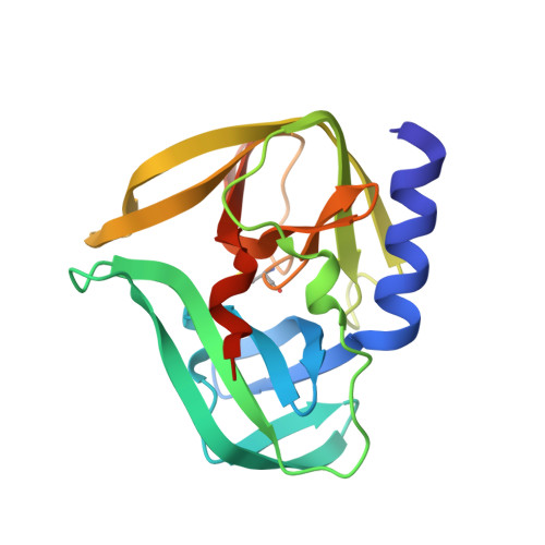
Structural Basis for Antiviral Inhibition of the Main Protease, 3C, from Human Enterovirus 93.

Costenaro, L.Kaczmarska, Z.Arnan, C.Janowski, R.Coutard, B.Sola, M.Gorbalenya, A.E.Norder, H.Canard, B.Coll, M.

(2011) J Virol 85: 10764-10773

  • DOI: https://doi.org/10.1128/JVI.05062-11
  • Primary Citation Related Structures: 
    3Q3X, 3Q3Y, 3RUO

  • PubMed Abstract: 

    Members of the Enterovirus genus of the Picornaviridae family are abundant, with common human pathogens that belong to the rhinovirus (HRV) and enterovirus (EV) species, including diverse echo-, coxsackie- and polioviruses. They cause a wide spectrum of clinical manifestations ranging from asymptomatic to severe diseases with neurological and/or cardiac manifestations. Pandemic outbreaks of EVs may be accompanied by meningitis and/or paralysis and can be fatal. However, no effective prophylaxis or antiviral treatment against most EVs is available. The EV RNA genome directs the synthesis of a single polyprotein that is autocatalytically processed into mature proteins at Gln↓Gly cleavage sites by the 3C protease (3C(pro)), which has narrow, conserved substrate specificity. These cleavages are essential for virus replication, making 3C(pro) an excellent target for antivirus drug development. In this study, we report the first determination of the crystal structure of 3C(pro) from an enterovirus B, EV-93, a recently identified pathogen, alone and in complex with the anti-HRV molecules compound 1 (AG7404) and rupintrivir (AG7088) at resolutions of 1.9, 1.3, and 1.5 Å, respectively. The EV-93 3C(pro) adopts a chymotrypsin-like fold with a canonically configured oxyanion hole and a substrate binding pocket similar to that of rhino-, coxsackie- and poliovirus 3C proteases. We show that compound 1 and rupintrivir are both active against EV-93 in infected cells and inhibit the proteolytic activity of EV-93 3C(pro) in vitro. These results provide a framework for further structure-guided optimization of the tested compounds to produce antiviral drugs against a broad range of EV species.


  • Organizational Affiliation
    • Institute for Research in Biomedicine, Barcelona, Spain.

Macromolecule Content 

  • Total Structure Weight: 43.49 kDa 
  • Atom Count: 3,415 
  • Modeled Residue Count: 365 
  • Deposited Residue Count: 382 
  • Unique protein chains: 1

Macromolecules

Find similar proteins by:|  3D Structure
Entity ID: 1
MoleculeChains  Sequence LengthOrganismDetailsImage
HEVB EV93 3C protease
A, B
191Enterovirus BMutation(s): 0 
Gene Names: 3C
EC: 3.4.22.28
UniProt
Find proteins for Q5DSM6 (Enterovirus B)
Explore Q5DSM6 
Go to UniProtKB:  Q5DSM6
Entity Groups
Sequence Clusters30% Identity50% Identity70% Identity90% Identity95% Identity100% Identity
UniProt GroupQ5DSM6
Sequence Annotations
Expand
Reference Sequence

Small Molecules

Ligands 2 Unique
IDChains Name / Formula / InChI Key2D Diagram3D Interactions
GOL

Query on GOL


Download:Ideal Coordinates CCD File
C [auth A]
D [auth A]
E [auth A]
H [auth B]
I [auth B]
C [auth A],
D [auth A],
E [auth A],
H [auth B],
I [auth B],
J [auth B]
GLYCEROL
C3 H8 O3
PEDCQBHIVMGVHV-UHFFFAOYSA-N
MG

Query on MG


Download:Ideal Coordinates CCD File
F [auth B],
G [auth B]
MAGNESIUM ION
Mg
JLVVSXFLKOJNIY-UHFFFAOYSA-N
Modified Residues  1 Unique
IDChains TypeFormula2D DiagramParent
OCS
Query on OCS
A, B
L-PEPTIDE LINKINGC3 H7 N O5 SCYS

Experimental Data & Validation

Experimental Data

  • Method: X-RAY DIFFRACTION
  • Resolution: 1.90 Å
  • R-Value Free:  0.210 (Depositor), 0.211 (DCC) 
  • R-Value Work:  0.148 (Depositor), 0.158 (DCC) 
  • R-Value Observed: 0.151 (Depositor) 
Space Group: P 1 21 1
Unit Cell:
Length ( Å )Angle ( ˚ )
a = 39.072α = 90
b = 65.216β = 90.67
c = 66.355γ = 90
Software Package:
Software NamePurpose
MOSFLMdata reduction
SCALAdata scaling
PHASERphasing
REFMACrefinement
PDB_EXTRACTdata extraction
MxCuBEdata collection

Structure Validation

View Full Validation Report


Entry History 

Deposition Data

Revision History  (Full details and data files)

  • Version 1.0: 2011-09-07
    Type: Initial release
  • Version 1.1: 2011-10-05
    Changes: Database references
  • Version 1.2: 2023-09-13
    Changes: Data collection, Database references, Derived calculations, Refinement description
  • Version 1.3: 2023-12-06
    Changes: Data collection