3Q2O | pdb_00003q2o

Crystal Structure of purK: N5-carboxyaminoimidazole ribonucleotide synthetase


Experimental Data Snapshot

  • Method: X-RAY DIFFRACTION
  • Resolution: 1.96 Å
  • R-Value Free: 
    0.235 (Depositor), 0.234 (DCC) 
  • R-Value Work: 
    0.195 (Depositor), 0.196 (DCC) 
  • R-Value Observed: 
    0.197 (Depositor) 

Starting Model: experimental
View more details

wwPDB Validation 3D Report Full Report

Validation slider image for 3Q2O

This is version 1.1 of the entry. See complete history

Literature

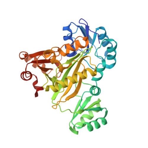
Structure of N(5)-carboxyaminoimidazole ribonucleotide synthase (PurK) from Bacillus anthracis.

Tuntland, M.L.Johnson, M.E.Fung, L.W.Santarsiero, B.D.

(2011) Acta Crystallogr D Biol Crystallogr 67: 870-874

  • DOI: https://doi.org/10.1107/S0907444911029210
  • Primary Citation Related Structures: 
    3Q2O

  • PubMed Abstract: 

    The apo structure of N5-carboxyaminoimidazole ribonucleotide synthase (PurK) from Bacillus anthracis (baPurK) with Mg2+ in the active site is reported at 1.96 Å resolution. PurK is an enzyme in the purine-biosynthetic pathway, unique to prokaryotes, that converts 5-aminoimidazole ribonucleotide to N5-carboxyaminoimidazole ribonucleotide and has been suggested as a potential antimicrobial drug target. Two interesting features of baPurK are a flexible B-loop (residues 149/150-157) that is in close contact with the active site and the binding of Mg2+ to the active site without additional ligands.


  • Organizational Affiliation
    • Department of Chemistry, University of Illinois at Chicago, Chicago, IL 60607, USA.

Macromolecule Content 

  • Total Structure Weight: 86.47 kDa 
  • Atom Count: 6,257 
  • Modeled Residue Count: 754 
  • Deposited Residue Count: 778 
  • Unique protein chains: 1

Macromolecules

Find similar proteins by:|  3D Structure
Entity ID: 1
MoleculeChains  Sequence LengthOrganismDetailsImage
Phosphoribosylaminoimidazole carboxylase, ATPase subunit
A, B
389Bacillus anthracis str. A0248Mutation(s): 0 
Gene Names: BAA_0344purK
EC: 4.1.1.21

Experimental Data & Validation

Experimental Data

  • Method: X-RAY DIFFRACTION
  • Resolution: 1.96 Å
  • R-Value Free:  0.235 (Depositor), 0.234 (DCC) 
  • R-Value Work:  0.195 (Depositor), 0.196 (DCC) 
  • R-Value Observed: 0.197 (Depositor) 
Space Group: P 21 21 21
Unit Cell:
Length ( Å )Angle ( ˚ )
a = 57.43α = 90
b = 82.445β = 90
c = 166.621γ = 90
Software Package:
Software NamePurpose
XSCALEdata scaling
PHASERphasing
REFMACrefinement
PDB_EXTRACTdata extraction
MAR345dtbdata collection
XDSdata reduction
XDSdata scaling

Structure Validation

View Full Validation Report


Entry History 

Deposition Data

Revision History  (Full details and data files)

  • Version 1.0: 2011-10-26
    Type: Initial release
  • Version 1.1: 2023-09-13
    Changes: Data collection, Database references, Derived calculations, Refinement description