


Crystal Structure of a pyridoxamine kinase from Yersinia pestis CO92
Brunzelle, J.S., Wawrzak, Z., Kudritska, M., Onopriyenko, O., Savchenko, A., Anderson, W.F., Center for Structural Genomics of Infectious Diseases (CSGID)To be published.
Experimental Data Snapshot
wwPDB Validation 3D Report Full Report

Entity ID: 1 | |||||
|---|---|---|---|---|---|
| Molecule | Chains | Sequence Length | Organism | Details | Image |
| Pyridoxamine kinase | 289 | Yersinia pestis CO92 | Mutation(s): 0 Gene Names: pdxY, y1967, YPO2368, YP_2154 EC: 2.7.1.35 | 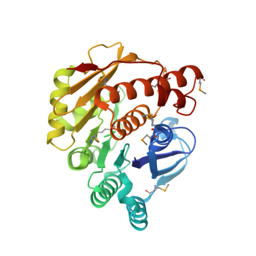 | |
UniProt | |||||
Entity Groups | |||||
| Sequence Clusters | 30% Identity50% Identity70% Identity90% Identity95% Identity100% Identity | ||||
| UniProt Group | Q7CIR8 | ||||
Sequence AnnotationsExpand | |||||
Reference Sequence | |||||
| Ligands 3 Unique | |||||
|---|---|---|---|---|---|
| ID | Chains | Name / Formula / InChI Key | 2D Diagram | 3D Interactions | |
| SO4 Download:Ideal Coordinates CCD File | D [auth A], J [auth B] | SULFATE ION O4 S QAOWNCQODCNURD-UHFFFAOYSA-L |  | ||
| BME Download:Ideal Coordinates CCD File | E [auth A] F [auth A] G [auth A] H [auth A] K [auth B] | BETA-MERCAPTOETHANOL C2 H6 O S DGVVWUTYPXICAM-UHFFFAOYSA-N |  | ||
| NA Download:Ideal Coordinates CCD File | C [auth A], I [auth B] | SODIUM ION Na FKNQFGJONOIPTF-UHFFFAOYSA-N |  | ||
| Modified Residues 1 Unique | |||||
|---|---|---|---|---|---|
| ID | Chains | Type | Formula | 2D Diagram | Parent |
| MSE Query on MSE | A, B | L-PEPTIDE LINKING | C5 H11 N O2 Se |  | MET |
| Length ( Å ) | Angle ( ˚ ) |
|---|---|
| a = 67.914 | α = 90 |
| b = 88.101 | β = 90 |
| c = 102.917 | γ = 90 |
| Software Name | Purpose |
|---|---|
| BLU-MAX | data collection |
| PHENIX | model building |
| BUSTER | refinement |
| HKL-2000 | data reduction |
| HKL-2000 | data scaling |
| PHENIX | phasing |


