3PYI | pdb_00003pyi

Structure of the N-terminal domain of C. elegans SAS-6


Experimental Data Snapshot

  • Method: X-RAY DIFFRACTION
  • Resolution: 2.10 Å
  • R-Value Free: 
    0.257 (Depositor), 0.258 (DCC) 
  • R-Value Work: 
    0.210 (Depositor), 0.208 (DCC) 
  • R-Value Observed: 
    0.212 (Depositor) 

wwPDB Validation 3D Report Full Report

Validation slider image for 3PYI

This is version 1.2 of the entry. See complete history

Literature

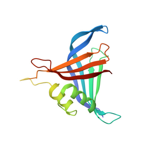
Structural basis of the 9-fold symmetry of centrioles.

Kitagawa, D.Vakonakis, I.Olieric, N.Hilbert, M.Keller, D.Olieric, V.Bortfeld, M.Erat, M.C.Fluckiger, I.Gonczy, P.Steinmetz, M.O.

(2011) Cell 144: 364-375

  • DOI: https://doi.org/10.1016/j.cell.2011.01.008
  • Primary Citation Related Structures: 
    3PYI, 3Q0X, 3Q0Y

  • PubMed Abstract: 

    The centriole, and the related basal body, is an ancient organelle characterized by a universal 9-fold radial symmetry and is critical for generating cilia, flagella, and centrosomes. The mechanisms directing centriole formation are incompletely understood and represent a fundamental open question in biology. Here, we demonstrate that the centriolar protein SAS-6 forms rod-shaped homodimers that interact through their N-terminal domains to form oligomers. We establish that such oligomerization is essential for centriole formation in C. elegans and human cells. We further generate a structural model of the related protein Bld12p from C. reinhardtii, in which nine homodimers assemble into a ring from which nine coiled-coil rods radiate outward. Moreover, we demonstrate that recombinant Bld12p self-assembles into structures akin to the central hub of the cartwheel, which serves as a scaffold for centriole formation. Overall, our findings establish a structural basis for the universal 9-fold symmetry of centrioles.


  • Organizational Affiliation
    • Swiss Institute for Experimental Cancer Research, School of Life Sciences, Swiss Federal Institute of Technology, EPFL, Lausanne, Switzerland.

Macromolecule Content 

  • Total Structure Weight: 39.85 kDa 
  • Atom Count: 2,547 
  • Modeled Residue Count: 285 
  • Deposited Residue Count: 340 
  • Unique protein chains: 1

Macromolecules

Find similar proteins by:|  3D Structure
Entity ID: 1
MoleculeChains  Sequence LengthOrganismDetailsImage
Spindle assembly abnormal protein 6A [auth B],
B [auth A]
170Caenorhabditis elegansMutation(s): 1 
Gene Names: sas-6Y45F10D.9
UniProt
Find proteins for O62479 (Caenorhabditis elegans)
Explore O62479 
Go to UniProtKB:  O62479
Entity Groups
Sequence Clusters30% Identity50% Identity70% Identity90% Identity95% Identity100% Identity
UniProt GroupO62479
Sequence Annotations
Expand
Reference Sequence

Experimental Data & Validation

Experimental Data

  • Method: X-RAY DIFFRACTION
  • Resolution: 2.10 Å
  • R-Value Free:  0.257 (Depositor), 0.258 (DCC) 
  • R-Value Work:  0.210 (Depositor), 0.208 (DCC) 
  • R-Value Observed: 0.212 (Depositor) 
Space Group: P 21 21 21
Unit Cell:
Length ( Å )Angle ( ˚ )
a = 70.273α = 90
b = 73.153β = 90
c = 79.596γ = 90
Software Package:
Software NamePurpose
MAR345dtbdata collection
PHENIXmodel building
PHENIXrefinement
XSCALEdata scaling
SCALAdata scaling
PHENIXphasing

Structure Validation

View Full Validation Report


Entry History 

Deposition Data

Revision History  (Full details and data files)

  • Version 1.0: 2011-02-09
    Type: Initial release
  • Version 1.1: 2011-07-13
    Changes: Version format compliance
  • Version 1.2: 2024-02-21
    Changes: Data collection, Database references, Derived calculations