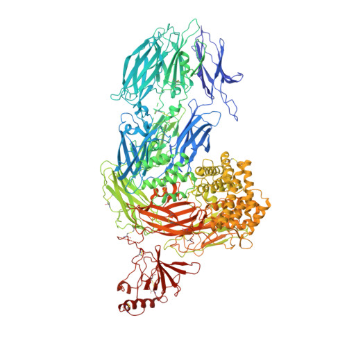
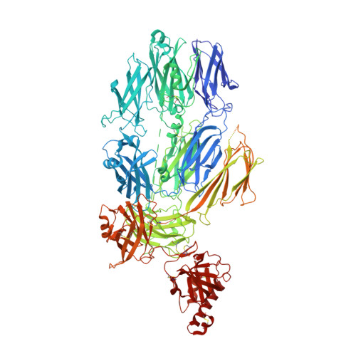
3PVM

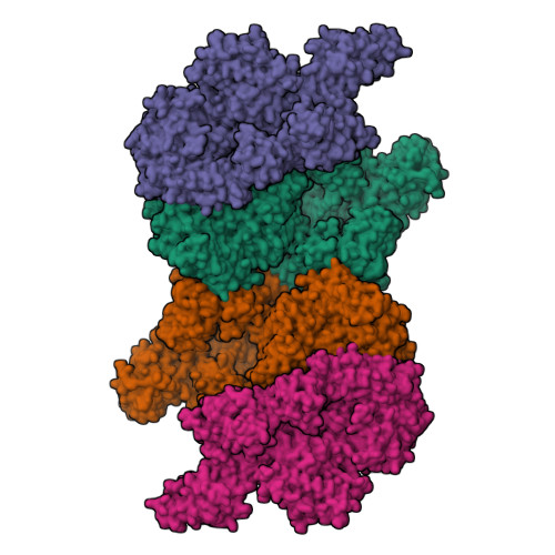
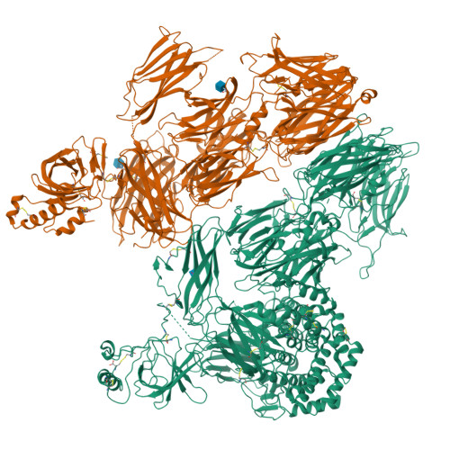
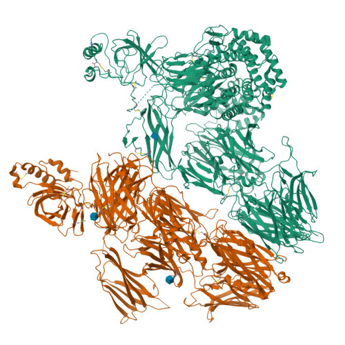
Structure of Complement C5 in Complex with CVF


Experimental Data Snapshot

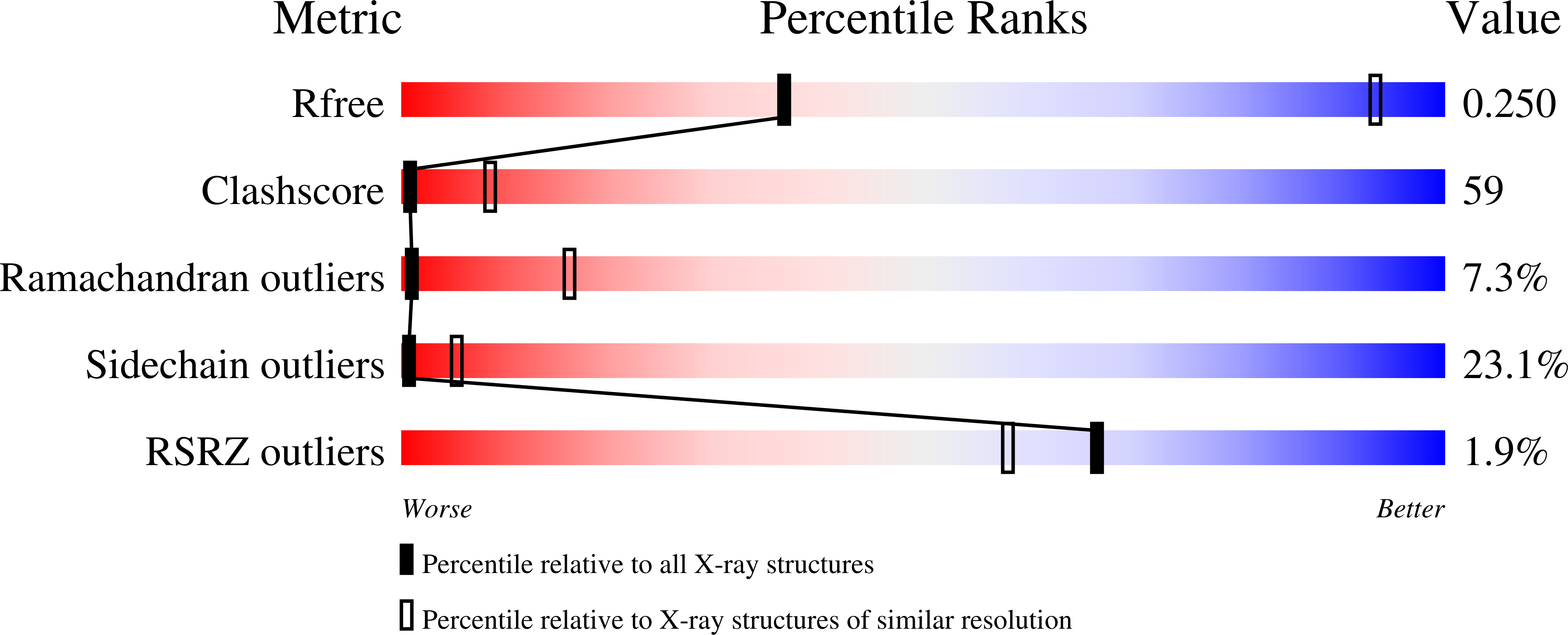
  • Method: X-RAY DIFFRACTION
  • Resolution: 4.30 Å
  • R-Value Free: 0.262 
  • R-Value Work: 0.233 
  • R-Value Observed: 0.234 

Starting Models: experimental
View more details

wwPDB Validation   3D Report Full Report


Ligand Structure Quality Assessment 


This is version 1.4 of the entry. See complete history


Literature

Substrate recognition by complement convertases revealed in the C5-cobra venom factor complex.

Laursen, N.S.Andersen, K.R.Braren, I.Spillner, E.Sottrup-Jensen, L.Andersen, G.R.

(2011) EMBO J 30: 606-616

  • DOI: https://doi.org/10.1038/emboj.2010.341
  • Primary Citation of Related Structures:  
    3PRX, 3PVM

  • PubMed Abstract: 

    Complement acts as a danger-sensing system in the innate immune system, and its activation initiates a strong inflammatory response and cleavage of the proteins C3 and C5 by proteolytic enzymes, the convertases. These contain a non-catalytic substrate contacting subunit (C3b or C4b) in complex with a protease subunit (Bb or C2a). We determined the crystal structures of the C3b homologue cobra venom factor (CVF) in complex with C5, and in complex with C5 and the inhibitor SSL7 at 4.3 Å resolution. The structures reveal a parallel two-point attachment between C5 and CVF, where the presence of SSL7 only slightly affects the C5-CVF interface, explaining the IgA dependence for SSL7-mediated inhibition of C5 cleavage. CVF functions as a relatively rigid binding scaffold inducing a conformational change in C5, which positions its cleavage site in proximity to the serine protease Bb. A general model for substrate recognition by the convertases is presented based on the C5-CVF and C3b-Bb-SCIN structures. Prior knowledge concerning interactions between the endogenous convertases and their substrates is rationalized by this model.


  • Organizational Affiliation

    Department of Molecular Biology, Aarhus University, Aarhus, Denmark.


Macromolecules
Find similar proteins by:  (by identity cutoff)  |  3D Structure
Entity ID: 1
MoleculeChains Sequence LengthOrganismDetailsImage
Complement C5
A, C
1,676Homo sapiensMutation(s): 0 
UniProt & NIH Common Fund Data Resources
Find proteins for P01031 (Homo sapiens)
Explore P01031 
Go to UniProtKB:  P01031
PHAROS:  P01031
GTEx:  ENSG00000106804 
Entity Groups  
Sequence Clusters30% Identity50% Identity70% Identity90% Identity95% Identity100% Identity
UniProt GroupP01031
Glycosylation
Glycosylation Sites: 1Go to GlyGen: P01031-1
Sequence Annotations
Expand
  • Reference Sequence
Find similar proteins by:  (by identity cutoff)  |  3D Structure
Entity ID: 2
MoleculeChains Sequence LengthOrganismDetailsImage
Cobra venom factor
B, D
1,642Naja kaouthiaMutation(s): 0 
UniProt
Find proteins for Q91132 (Naja kaouthia)
Explore Q91132 
Go to UniProtKB:  Q91132
Entity Groups  
Sequence Clusters30% Identity50% Identity70% Identity90% Identity95% Identity100% Identity
UniProt GroupQ91132
Glycosylation
Glycosylation Sites: 2
Sequence Annotations
Expand
  • Reference Sequence
Experimental Data & Validation

Experimental Data

  • Method: X-RAY DIFFRACTION
  • Resolution: 4.30 Å
  • R-Value Free: 0.262 
  • R-Value Work: 0.233 
  • R-Value Observed: 0.234 
  • Space Group: P 2 2 21
Unit Cell:
Length ( Å )Angle ( ˚ )
a = 176.52α = 90
b = 179.2β = 90
c = 389.69γ = 90
Software Package:
Software NamePurpose
PHENIXrefinement
XDSdata reduction
XSCALEdata scaling
PHASERphasing

Structure Validation

View Full Validation Report


Ligand Structure Quality Assessment 


Entry History 

Deposition Data

Revision History  (Full details and data files)

  • Version 1.0: 2011-01-19
    Type: Initial release
  • Version 1.1: 2011-07-13
    Changes: Version format compliance
  • Version 1.2: 2014-01-22
    Changes: Refinement description
  • Version 1.3: 2020-07-29
    Type: Remediation
    Reason: Carbohydrate remediation
    Changes: Data collection, Derived calculations, Structure summary
  • Version 1.4: 2023-09-06
    Changes: Data collection, Database references, Refinement description, Structure summary