3PSL | pdb_00003psl

Fine-tuning the stimulation of MLL1 methyltransferase activity by a histone H3 based peptide mimetic


Experimental Data Snapshot

  • Method: X-RAY DIFFRACTION
  • Resolution: 1.70 Å
  • R-Value Free: 
    0.205 (Depositor), 0.214 (DCC) 
  • R-Value Work: 
    0.155 (Depositor), 0.162 (DCC) 
  • R-Value Observed: 
    0.157 (Depositor) 

Starting Model: experimental
View more details

wwPDB Validation 3D Report Full Report

Validation slider image for 3PSL

This is version 1.4 of the entry. See complete history

Literature

Fine-tuning the stimulation of MLL1 methyltransferase activity by a histone H3-based peptide mimetic.

Avdic, V.Zhang, P.Lanouette, S.Voronova, A.Skerjanc, I.Couture, J.F.

(2011) FASEB J 25: 960-967

  • DOI: https://doi.org/10.1096/fj.10-171959
  • Primary Citation Related Structures: 
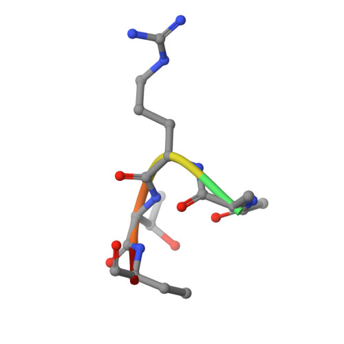
    3PSL

  • PubMed Abstract: 

    The SET1 family of methyltransferases carries out the bulk of histone H3 Lys-4 methylation in vivo. One of the common features of this family is the regulation of their methyltransferase activity by a tripartite complex composed of WDR5, RbBP5, and Ash2L. To selectively probe the role of the SET1 family of methyltransferases, we have developed a library of histone H3 peptide mimetics and report herein the characterization of an Nα acetylated form of histone H3 peptide (NαH3). Binding and inhibition studies reveal that the addition of an acetyl moiety to the N terminus of histone H3 significantly enhances its binding to WDR5 and prevents the stimulation of MLL1 methyltransferase activity by the WDR5-RbBP5-Ash2L complex. The crystal structure of NαH3 in complex with WDR5 reveals that a high-affinity hydrophobic pocket accommodates the binding of the acetyl moiety. These results provide the structural basis to control WDR5-RbBP5-Ash2L-MLL1 activity and a tool to manipulate stem cell differentiation programs.


  • Organizational Affiliation
    • University of Ottawa, Ottawa Institute of Systems Biology, 451 Smyth Rd., Roger Guindon Hall, Ottawa, ON K1H 8M5, Canada.

Macromolecule Content 

  • Total Structure Weight: 70.85 kDa 
  • Atom Count: 5,019 
  • Modeled Residue Count: 606 
  • Deposited Residue Count: 648 
  • Unique protein chains: 2

Macromolecules

Find similar proteins by:|  3D Structure
Entity ID: 1
MoleculeChains  Sequence LengthOrganismDetailsImage
WD repeat-containing protein 5
A, B
318Homo sapiensMutation(s): 0 
Gene Names: WDR5BIG3
UniProt & NIH Common Fund Data Resources
Find proteins for P61964 (Homo sapiens)
Explore P61964 
Go to UniProtKB:  P61964
PHAROS:  P61964
GTEx:  ENSG00000196363 
Entity Groups
Sequence Clusters30% Identity50% Identity70% Identity90% Identity95% Identity100% Identity
UniProt GroupP61964
Sequence Annotations
Expand
Reference Sequence
Find similar proteins by:  Sequence   |   3D Structure  
Entity ID: 2
MoleculeChains  Sequence LengthOrganismDetailsImage
N-alpha acetylated form of histone H3
C, D
6N/AMutation(s): 0 

Experimental Data & Validation

Experimental Data

  • Method: X-RAY DIFFRACTION
  • Resolution: 1.70 Å
  • R-Value Free:  0.205 (Depositor), 0.214 (DCC) 
  • R-Value Work:  0.155 (Depositor), 0.162 (DCC) 
  • R-Value Observed: 0.157 (Depositor) 
Space Group: P 1
Unit Cell:
Length ( Å )Angle ( ˚ )
a = 45.947α = 98.79
b = 48.614β = 90.95
c = 63.308γ = 117.48
Software Package:
Software NamePurpose
Blu-Icedata collection
MOLREPphasing
REFMACrefinement
HKL-2000data reduction
HKL-2000data scaling

Structure Validation

View Full Validation Report


Entry History 

Deposition Data

Revision History  (Full details and data files)

  • Version 1.0: 2010-12-22
    Type: Initial release
  • Version 1.1: 2011-07-13
    Changes: Version format compliance
  • Version 1.2: 2011-08-24
    Changes: Database references
  • Version 1.3: 2023-09-06
    Changes: Data collection, Database references, Derived calculations, Refinement description
  • Version 1.4: 2024-11-27
    Changes: Structure summary