3PQV | pdb_00003pqv

Cyclase homolog


Experimental Data Snapshot

  • Method: X-RAY DIFFRACTION
  • Resolution: 2.61 Å
  • R-Value Free: 
    0.259 (Depositor), 0.253 (DCC) 
  • R-Value Work: 
    0.204 (Depositor), 0.200 (DCC) 
  • R-Value Observed: 
    0.207 (Depositor) 

wwPDB Validation 3D Report Full Report

Validation slider image for 3PQV

This is version 1.2 of the entry. See complete history

Literature

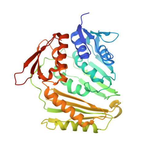
Crystal structure of Rcl1, an essential component of the eukaryal pre-rRNA processosome implicated in 18s rRNA biogenesis.

Tanaka, N.Smith, P.Shuman, S.

(2011) RNA 17: 595-602

  • DOI: https://doi.org/10.1261/rna.2571811
  • Primary Citation Related Structures: 
    3PQV

  • PubMed Abstract: 

    Rcl1 is an essential nucleolar protein required for U3 snoRNA-guided pre-rRNA processing at sites flanking the 18S rRNA sequence. A potential catalytic role for Rcl1 during pre-rRNA cleavage has been suggested based on its primary structure similarity to RNA 3'-terminal phosphate cyclase (Rtc) enzymes, which perform nucleotidyl transfer and phosphoryl transfer reactions at RNA ends. Here, we report the 2.6 Å crystal structure of a biologically active yeast Rcl1, which illuminates its modular 4-domain architecture and overall homology with RNA cyclases while revealing numerous local differences that account for why Rtcs possess metal-dependent adenylyltransferase activity and Rcls do not. A conserved oxyanion-binding site in Rcl1 was highlighted for possible catalytic or RNA-binding functions. However, the benign effects of mutations in and around the anion site on Rcl1 activity in vivo militate against such a role.


  • Organizational Affiliation
    • Molecular Biology Program, Sloan-Kettering Institute, New York, New York 10065, USA.

Macromolecule Content 

  • Total Structure Weight: 161.45 kDa 
  • Atom Count: 11,342 
  • Modeled Residue Count: 1,409 
  • Deposited Residue Count: 1,460 
  • Unique protein chains: 1

Macromolecules

Find similar proteins by:|  3D Structure
Entity ID: 1
MoleculeChains  Sequence LengthOrganismDetailsImage
Rcl1 protein
A, B, C, D
365Kluyveromyces lactisMutation(s): 0 
Gene Names: KLLA0C05984g
UniProt
Find proteins for Q6CUC5 (Kluyveromyces lactis (strain ATCC 8585 / CBS 2359 / DSM 70799 / NBRC 1267 / NRRL Y-1140 / WM37))
Explore Q6CUC5 
Go to UniProtKB:  Q6CUC5
Entity Groups
Sequence Clusters30% Identity50% Identity70% Identity90% Identity95% Identity100% Identity
UniProt GroupQ6CUC5
Sequence Annotations
Expand
Reference Sequence

Small Molecules

Ligands 3 Unique
IDChains Name / Formula / InChI Key2D Diagram3D Interactions
TAR

Query on TAR


Download:Ideal Coordinates CCD File
E [auth A],
G [auth B],
I [auth C],
J [auth D],
K [auth D]
D(-)-TARTARIC ACID
C4 H6 O6
FEWJPZIEWOKRBE-LWMBPPNESA-N
PEG

Query on PEG


Download:Ideal Coordinates CCD File
F [auth A],
H [auth B]
DI(HYDROXYETHYL)ETHER
C4 H10 O3
MTHSVFCYNBDYFN-UHFFFAOYSA-N
GOL

Query on GOL


Download:Ideal Coordinates CCD File
L [auth D]GLYCEROL
C3 H8 O3
PEDCQBHIVMGVHV-UHFFFAOYSA-N

Experimental Data & Validation

Experimental Data

  • Method: X-RAY DIFFRACTION
  • Resolution: 2.61 Å
  • R-Value Free:  0.259 (Depositor), 0.253 (DCC) 
  • R-Value Work:  0.204 (Depositor), 0.200 (DCC) 
  • R-Value Observed: 0.207 (Depositor) 
Space Group: P 21 21 21
Unit Cell:
Length ( Å )Angle ( ˚ )
a = 59.317α = 90
b = 138.821β = 90
c = 228.945γ = 90
Software Package:
Software NamePurpose
HKL-2000data collection
PHENIXmodel building
PHENIXrefinement
HKL-2000data reduction
SCALEPACKdata scaling
PHENIXphasing

Structure Validation

View Full Validation Report


Entry History 

Deposition Data

Revision History  (Full details and data files)

  • Version 1.0: 2011-04-13
    Type: Initial release
  • Version 1.1: 2011-07-13
    Changes: Version format compliance
  • Version 1.2: 2024-02-21
    Changes: Data collection, Database references, Derived calculations