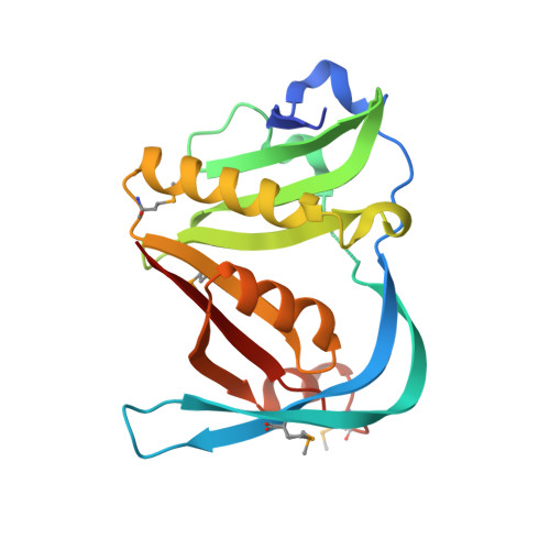
1.6 Angstrom resolution crystal structure of putative streptothricin acetyltransferase from Bacillus anthracis str. Ames in complex with acetyl coenzyme A
Halavaty, A.S., Wawrzak, Z., Onopriyenko, O., Edwards, A., Savchenko, A., Peterson, S., Anderson, W.F., Center for Structural Genomics of Infectious Diseases (CSGID)To be published.