3POY | pdb_00003poy

Crystal Structure of the alpha-Neurexin-1 ectodomain, LNS 2-6


Experimental Data Snapshot

  • Method: X-RAY DIFFRACTION
  • Resolution: 3.02 Å
  • R-Value Free: 
    0.269 (Depositor), 0.262 (DCC) 
  • R-Value Work: 
    0.214 (Depositor), 0.216 (DCC) 
  • R-Value Observed: 
    0.216 (Depositor) 

Starting Models: experimental
View more details

wwPDB Validation 3D Report Full Report

Validation slider image for 3POY

Ligand Structure Quality Assessment 


This is version 2.3 of the entry. See complete history

Literature

The crystal structure of the alpha-neurexin-1 extracellular region reveals a hinge point for mediating synaptic adhesion and function.

Miller, M.T.Mileni, M.Comoletti, D.Stevens, R.C.Harel, M.Taylor, P.

(2011) Structure 19: 767-778

  • DOI: https://doi.org/10.1016/j.str.2011.03.011
  • Primary Citation Related Structures: 
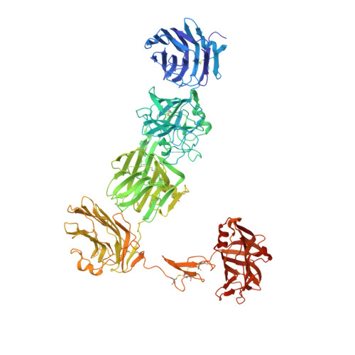
    3POY

  • PubMed Abstract: 

    α- and β-neurexins (NRXNs) are transmembrane cell adhesion proteins that localize to presynaptic membranes in neurons and interact with the postsynaptic neuroligins (NLGNs). Their gene mutations are associated with the autism spectrum disorders. The extracellular region of α-NRXNs, containing nine independently folded domains, has structural complexity and unique functional characteristics, distinguishing it from the smaller β-NRXNs. We have solved the X-ray crystal structure of seven contiguous domains of the α-NRXN-1 extracellular region at 3.0 Å resolution. The structure reveals an arrangement where the N-terminal five domains adopt a more rigid linear conformation and the two C-terminal domains form a separate arm connected by a flexible hinge. In an extended conformation the molecule is suitably configured to accommodate a bound NLGN molecule, as supported by structural comparison and surface plasmon resonance. These studies provide the structural basis for a multifunctional synaptic adhesion complex mediated by α-NRXN-1.


  • Organizational Affiliation
    • Department of Pharmacology, Skaggs School of Pharmacy and Pharmaceutical Sciences, University of California, San Diego, La Jolla, CA 92093, USA. m4miller@ucsd.edu

Macromolecule Content 

  • Total Structure Weight: 113.36 kDa 
  • Atom Count: 7,878 
  • Modeled Residue Count: 1,005 
  • Deposited Residue Count: 1,019 
  • Unique protein chains: 1

Macromolecules

Find similar proteins by:|  3D Structure
Entity ID: 1
MoleculeChains  Sequence LengthOrganismDetailsImage
Neurexin-1-alpha1,019Bos taurusMutation(s): 2 
Gene Names: NRXN1
UniProt
Find proteins for Q28146 (Bos taurus)
Explore Q28146 
Go to UniProtKB:  Q28146
Entity Groups
Sequence Clusters30% Identity50% Identity70% Identity90% Identity95% Identity100% Identity
UniProt GroupQ28146
Glycosylation
Glycosylation Sites: 2
Sequence Annotations
Expand
Reference Sequence

Oligosaccharides

Help  
Entity ID: 2
MoleculeChains Length2D Diagram GlycosylationD Interactions
beta-D-mannopyranose-(1-4)-2-acetamido-2-deoxy-beta-D-glucopyranose-(1-4)-2-acetamido-2-deoxy-beta-D-glucopyranose
B
3N-Glycosylation
Glycosylation Resources
GlyTouCan: G15407YE
GlyCosmos: G15407YE
GlyGen: G15407YE

Small Molecules

Ligands 1 Unique
IDChains Name / Formula / InChI Key2D Diagram3D Interactions
BGC

Query on BGC


Download:Ideal Coordinates CCD File
C [auth A]beta-D-glucopyranose
C6 H12 O6
WQZGKKKJIJFFOK-VFUOTHLCSA-N

Experimental Data & Validation

Experimental Data

  • Method: X-RAY DIFFRACTION
  • Resolution: 3.02 Å
  • R-Value Free:  0.269 (Depositor), 0.262 (DCC) 
  • R-Value Work:  0.214 (Depositor), 0.216 (DCC) 
  • R-Value Observed: 0.216 (Depositor) 
Space Group: C 1 2 1
Unit Cell:
Length ( Å )Angle ( ˚ )
a = 199.74α = 90
b = 61.24β = 121.21
c = 155.58γ = 90
Software Package:
Software NamePurpose
XDSdata scaling
PHASERphasing
REFMACrefinement
XDSdata reduction
XSCALEdata scaling

Structure Validation

View Full Validation Report


Ligand Structure Quality Assessment 


Entry History 

Deposition Data

Revision History  (Full details and data files)

  • Version 1.0: 2011-06-08
    Type: Initial release
  • Version 1.1: 2011-07-13
    Changes: Version format compliance
  • Version 1.2: 2013-06-19
    Changes: Database references
  • Version 2.0: 2020-07-29
    Type: Remediation
    Reason: Carbohydrate remediation
    Changes: Atomic model, Data collection, Database references, Derived calculations, Structure summary
  • Version 2.1: 2021-03-31
    Changes: Source and taxonomy, Structure summary
  • Version 2.2: 2023-09-06
    Changes: Data collection, Database references, Refinement description
  • Version 2.3: 2024-11-27
    Changes: Structure summary