3POW

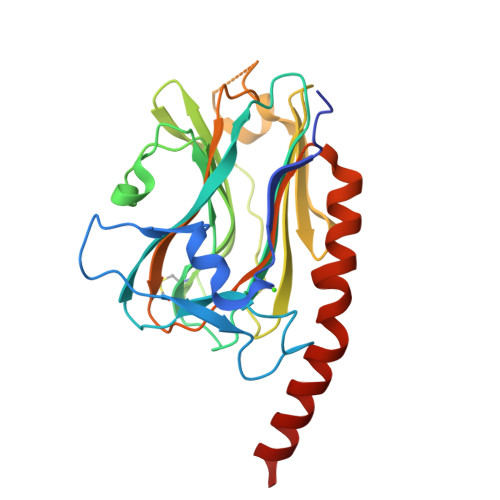
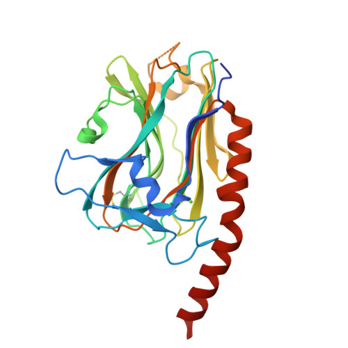
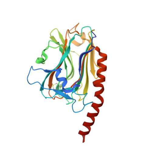
Crystal structure of the globular domain of human calreticulin


Experimental Data Snapshot

  • Method: X-RAY DIFFRACTION
  • Resolution: 1.55 Å
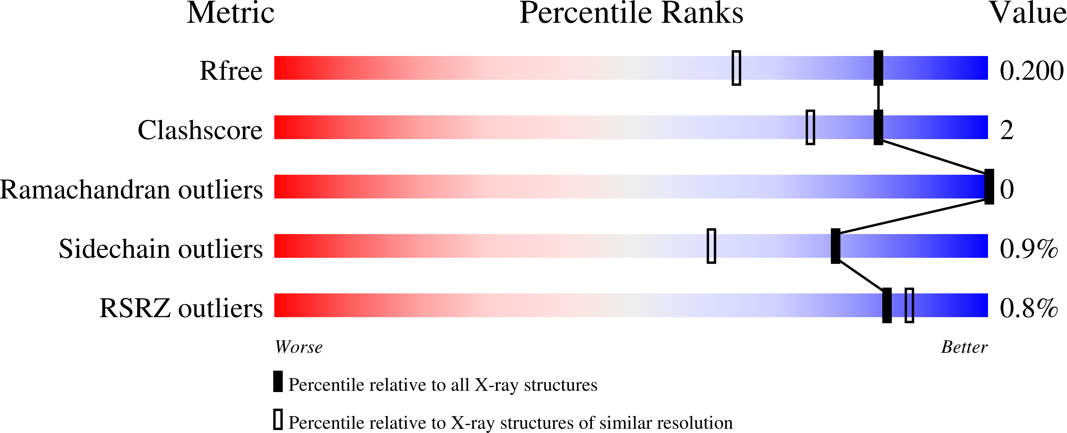
  • R-Value Free: 0.191 
  • R-Value Work: 0.176 
  • R-Value Observed: 0.177 

Starting Model: experimental
View more details

wwPDB Validation   3D Report Full Report


This is version 1.4 of the entry. See complete history


Literature

X-ray structure of the human calreticulin globular domain reveals a Peptide-binding area and suggests a multi-molecular mechanism

Chouquet, A.Paidassi, H.Ling, W.L.Frachet, P.Houen, G.Arlaud, G.J.Gaboriaud, C.

(2011) PLoS One 6: e17886-e17886

  • DOI: https://doi.org/10.1371/journal.pone.0017886
  • Primary Citation of Related Structures:  
    3POS, 3POW

  • PubMed Abstract: 

    In the endoplasmic reticulum, calreticulin acts as a chaperone and a Ca(2+)-signalling protein. At the cell surface, it mediates numerous important biological effects. The crystal structure of the human calreticulin globular domain was solved at 1.55 Å resolution. Interactions of the flexible N-terminal extension with the edge of the lectin site are consistently observed, revealing a hitherto unidentified peptide-binding site. A calreticulin molecular zipper, observed in all crystal lattices, could further extend this site by creating a binding cavity lined by hydrophobic residues. These data thus provide a first structural insight into the lectin-independent binding properties of calreticulin and suggest new working hypotheses, including that of a multi-molecular mechanism.


  • Organizational Affiliation

    Institut de Biologie Structurale Jean-Pierre Ebel, CEA, Grenoble, France.


Macromolecules
Find similar proteins by:  (by identity cutoff)  |  3D Structure
Entity ID: 1
MoleculeChains Sequence LengthOrganismDetailsImage
calreticulin265Homo sapiensMutation(s): 0 
Gene Names: CALRCRTC
UniProt & NIH Common Fund Data Resources
Find proteins for P27797 (Homo sapiens)
Explore P27797 
Go to UniProtKB:  P27797
PHAROS:  P27797
GTEx:  ENSG00000179218 
Entity Groups  
Sequence Clusters30% Identity50% Identity70% Identity90% Identity95% Identity100% Identity
UniProt GroupP27797
Sequence Annotations
Expand
  • Reference Sequence
Small Molecules
Ligands 1 Unique
IDChains Name / Formula / InChI Key2D Diagram3D Interactions
CA
Query on CA

Download Ideal Coordinates CCD File 
B [auth A]CALCIUM ION
Ca
BHPQYMZQTOCNFJ-UHFFFAOYSA-N
Experimental Data & Validation

Experimental Data

  • Method: X-RAY DIFFRACTION
  • Resolution: 1.55 Å
  • R-Value Free: 0.191 
  • R-Value Work: 0.176 
  • R-Value Observed: 0.177 
  • Space Group: P 21 21 21
Unit Cell:
Length ( Å )Angle ( ˚ )
a = 42.89α = 90
b = 70.39β = 90
c = 91.82γ = 90
Software Package:
Software NamePurpose
XSCALEdata scaling
BUSTER-TNTrefinement
PDB_EXTRACTdata extraction
XDSdata scaling
XDSdata reduction
PHASERphasing
BUSTERrefinement

Structure Validation

View Full Validation Report


Entry History 

Deposition Data

Revision History  (Full details and data files)

  • Version 1.0: 2011-03-09
    Type: Initial release
  • Version 1.1: 2011-07-13
    Changes: Version format compliance
  • Version 1.2: 2017-08-09
    Changes: Data collection, Refinement description, Source and taxonomy
  • Version 1.3: 2023-11-01
    Changes: Data collection, Database references, Derived calculations, Refinement description
  • Version 1.4: 2024-10-30
    Changes: Structure summary