3POV | pdb_00003pov

Crystal structure of a SOX-DNA complex


Experimental Data Snapshot

  • Method: X-RAY DIFFRACTION
  • Resolution: 2.50 Å
  • R-Value Free: 
    0.264 (Depositor), 0.258 (DCC) 
  • R-Value Work: 
    0.206 (Depositor), 0.200 (DCC) 
  • R-Value Observed: 
    0.209 (Depositor) 

Starting Model: experimental
View more details

wwPDB Validation 3D Report Full Report

Validation slider image for 3POV

This is version 1.1 of the entry. See complete history

Literature

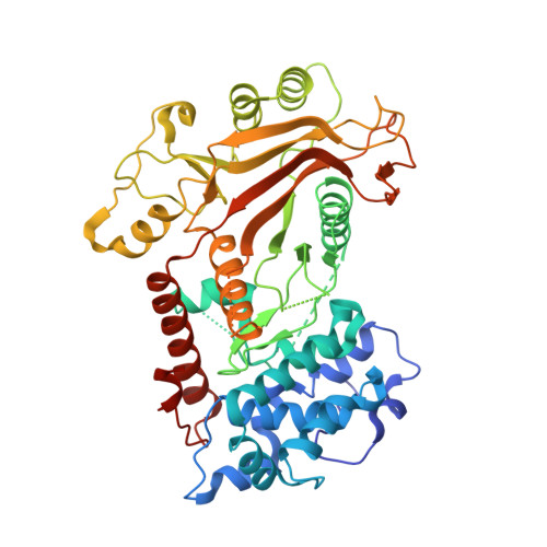
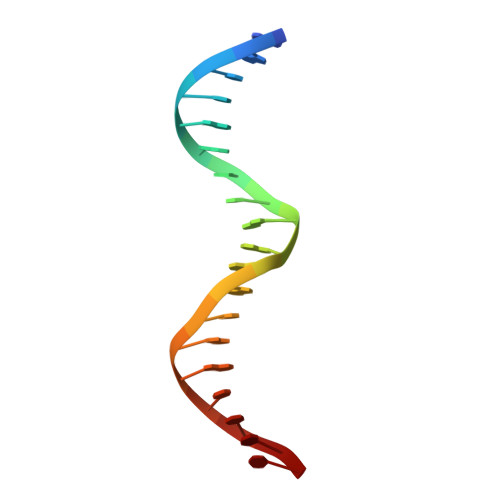
Crystal structure of a KSHV-SOX-DNA complex: insights into the molecular mechanisms underlying DNase activity and host shutoff

Bagneris, C.Briggs, L.C.Savva, R.Ebrahimi, B.Barrett, T.E.

(2011) Nucleic Acids Res 39: 5744-5756

  • DOI: https://doi.org/10.1093/nar/gkr111
  • Primary Citation Related Structures: 
    3POV

  • PubMed Abstract: 

    The early lytic phase of Kaposi's sarcoma herpesvirus infection is characterized by viral replication and the global degradation (shutoff) of host mRNA. Key to both activities is the virally encoded alkaline exonuclease KSHV SOX. While the DNase activity of KSHV SOX is required for the resolution of viral genomic DNA as a precursor to encapsidation, its exact involvement in host shutoff remains to be determined. We present the first crystal structure of a KSHV SOX-DNA complex that has illuminated the catalytic mechanism underpinning both its endo and exonuclease activities. We further illustrate that KSHV SOX, similar to its Epstein-Barr virus homologue, has an intrinsic RNase activity in vitro that although an element of host shutoff, cannot solely account for the phenomenon.


  • Organizational Affiliation
    • Institute of Structural and Molecular Biology, Crystallography, Department of Biological Sciences, Birkbeck College, Malet Street, London WC1E 7HX , UK.

Macromolecule Content 

  • Total Structure Weight: 67.03 kDa 
  • Atom Count: 4,373 
  • Modeled Residue Count: 483 
  • Deposited Residue Count: 526 
  • Unique protein chains: 1
  • Unique nucleic acid chains: 2

Macromolecules


Find similar proteins by:|  3D Structure
Entity ID: 1
MoleculeChains  Sequence LengthOrganismDetailsImage
ORF 37488Human herpesvirus 8 type MMutation(s): 0 
Gene Names: orf37
UniProt
Find proteins for P88925 (Human herpesvirus 8 type M)
Explore P88925 
Go to UniProtKB:  P88925
Entity Groups
Sequence Clusters30% Identity50% Identity70% Identity90% Identity95% Identity100% Identity
UniProt GroupP88925
Sequence Annotations
Expand
Reference Sequence
Find similar nucleic acids by:  Sequence
Entity ID: 2
MoleculeChains LengthOrganismImage
DNA (5'-D(*GP*GP*GP*AP*TP*CP*CP*TP*CP*CP*CP*AP*GP*TP*CP*GP*AP*CP*C)-3')B [auth C]19N/A
Sequence Annotations
Expand
Reference Sequence
Find similar nucleic acids by:  Sequence
Entity ID: 3
MoleculeChains LengthOrganismImage
DNA (5'-D(*GP*GP*TP*CP*GP*AP*CP*TP*AP*GP*GP*AP*GP*GP*AP*TP*CP*CP*C)-3')C [auth D]19N/A
Sequence Annotations
Expand
Reference Sequence

Experimental Data & Validation

Experimental Data

  • Method: X-RAY DIFFRACTION
  • Resolution: 2.50 Å
  • R-Value Free:  0.264 (Depositor), 0.258 (DCC) 
  • R-Value Work:  0.206 (Depositor), 0.200 (DCC) 
  • R-Value Observed: 0.209 (Depositor) 
Space Group: P 21 21 21
Unit Cell:
Length ( Å )Angle ( ˚ )
a = 54.52α = 90
b = 67.66β = 90
c = 174.56γ = 90
Software Package:
Software NamePurpose
DNAdata collection
MOLREPphasing
PHENIXrefinement
MOSFLMdata reduction
SCALAdata scaling

Structure Validation

View Full Validation Report


Entry History 

Deposition Data

Revision History  (Full details and data files)

  • Version 1.0: 2011-09-21
    Type: Initial release
  • Version 1.1: 2023-11-01
    Changes: Data collection, Database references, Derived calculations, Refinement description