3PND | pdb_00003pnd

FAD binding by ApbE protein from Salmonella enterica: a new class of FAD binding proteins


Experimental Data Snapshot

  • Method: X-RAY DIFFRACTION
  • Resolution: 2.75 Å
  • R-Value Free: 
    0.254 (Depositor), 0.258 (DCC) 
  • R-Value Work: 
    0.202 (Depositor), 0.205 (DCC) 
  • R-Value Observed: 
    0.205 (Depositor) 

Starting Model: experimental
View more details

wwPDB Validation 3D Report Full Report

Validation slider image for 3PND

Ligand Structure Quality Assessment 


This is version 1.3 of the entry. See complete history

Literature

FAD binding by ApbE protein from Salmonella enterica: a new class of FAD-binding proteins.

Boyd, J.M.Endrizzi, J.A.Hamilton, T.L.Christopherson, M.R.Mulder, D.W.Downs, D.M.Peters, J.W.

(2011) J Bacteriol 193: 887-895

  • DOI: https://doi.org/10.1128/JB.00730-10
  • Primary Citation Related Structures: 
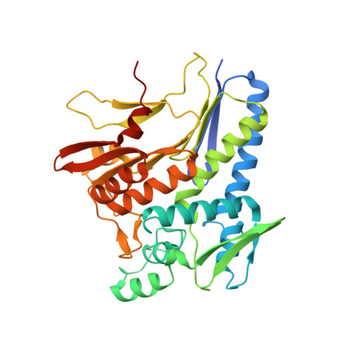
    3PND

  • PubMed Abstract: 

    The periplasmic protein ApbE was identified through the analysis of several mutants defective in thiamine biosynthesis and was implicated as having a role in iron-sulfur cluster biosynthesis or repair. While mutations in apbE cause decreased activity of several iron-sulfur enzymes in vivo, the specific role of ApbE remains unknown. Members of the AbpE family include NosX and RnfF, which have been implicated in oxidation-reduction associated with nitrous oxide and nitrogen metabolism, respectively. In this work, we show that ApbE binds one FAD molecule per monomeric unit. The structure of ApbE in the presence of bound FAD reveals a new FAD-binding motif. Protein variants that are nonfunctional in vivo were generated by random and targeted mutagenesis. Each variant was substituted in the environment of the FAD and analyzed for FAD binding after reconstitution. The variant that altered a key tyrosine residue involved in FAD binding prevented reconstitution of the protein.


  • Organizational Affiliation
    • Department of Bacteriology, University of Wisconsin, Madison, WI 53706, USA.

Macromolecule Content 

  • Total Structure Weight: 155.31 kDa 
  • Atom Count: 10,442 
  • Modeled Residue Count: 1,259 
  • Deposited Residue Count: 1,348 
  • Unique protein chains: 1

Macromolecules

Find similar proteins by:|  3D Structure
Entity ID: 1
MoleculeChains  Sequence LengthOrganismDetailsImage
Thiamine biosynthesis lipoprotein ApbE
A, B, C, D
337Salmonella enterica subsp. enterica serovar TyphimuriumMutation(s): 0 
Gene Names: apbESTM2266
EC: 2.7.1.180
UniProt
Find proteins for P41780 (Salmonella typhimurium (strain LT2 / SGSC1412 / ATCC 700720))
Explore P41780 
Go to UniProtKB:  P41780
Entity Groups
Sequence Clusters30% Identity50% Identity70% Identity90% Identity95% Identity100% Identity
UniProt GroupP41780
Sequence Annotations
Expand
Reference Sequence

Small Molecules

Ligands 2 Unique
IDChains Name / Formula / InChI Key2D Diagram3D Interactions
FAD

Query on FAD


Download:Ideal Coordinates CCD File
CA [auth B],
L [auth A],
NA [auth C],
WA [auth D]
FLAVIN-ADENINE DINUCLEOTIDE
C27 H33 N9 O15 P2
VWWQXMAJTJZDQX-UYBVJOGSSA-N
SO4

Query on SO4


Download:Ideal Coordinates CCD File
AA [auth B]
BA [auth B]
DA [auth B]
E [auth A]
EA [auth B]
AA [auth B],
BA [auth B],
DA [auth B],
E [auth A],
EA [auth B],
F [auth A],
FA [auth C],
G [auth A],
GA [auth C],
H [auth A],
HA [auth C],
I [auth A],
IA [auth C],
J [auth A],
JA [auth C],
K [auth A],
KA [auth C],
LA [auth C],
M [auth A],
MA [auth C],
N [auth A],
O [auth A],
OA [auth C],
P [auth A],
PA [auth C],
Q [auth A],
QA [auth C],
R [auth A],
RA [auth D],
S [auth B],
SA [auth D],
T [auth B],
TA [auth D],
U [auth B],
UA [auth D],
V [auth B],
VA [auth D],
W [auth B],
X [auth B],
XA [auth D],
Y [auth B],
YA [auth D],
Z [auth B],
ZA [auth D]
SULFATE ION
O4 S
QAOWNCQODCNURD-UHFFFAOYSA-L

Experimental Data & Validation

Experimental Data

  • Method: X-RAY DIFFRACTION
  • Resolution: 2.75 Å
  • R-Value Free:  0.254 (Depositor), 0.258 (DCC) 
  • R-Value Work:  0.202 (Depositor), 0.205 (DCC) 
  • R-Value Observed: 0.205 (Depositor) 
Space Group: P 21 21 21
Unit Cell:
Length ( Å )Angle ( ˚ )
a = 65.103α = 90
b = 120.85β = 90
c = 212.058γ = 90
Software Package:
Software NamePurpose
MOSFLMdata reduction
SCALAdata scaling
REFMACrefinement
PDB_EXTRACTdata extraction

Structure Validation

View Full Validation Report


Ligand Structure Quality Assessment 


Entry History 

Deposition Data

Revision History  (Full details and data files)

  • Version 1.0: 2011-01-05
    Type: Initial release
  • Version 1.1: 2011-07-13
    Changes: Version format compliance
  • Version 1.2: 2011-11-02
    Changes: Database references
  • Version 1.3: 2023-09-06
    Changes: Data collection, Database references, Derived calculations, Refinement description