


Crystal structure of a Putative dehydrogenase (RPA1076) from Rhodopseudomonas palustris CGA009 at 2.57 A resolution
Joint Center for Structural Genomics (JCSG)To be published.
Experimental Data Snapshot
Entity ID: 1 | |||||
|---|---|---|---|---|---|
| Molecule | Chains | Sequence Length | Organism | Details | Image |
| Putative oxidoreductase | 476 | Rhodopseudomonas palustris | Mutation(s): 0 Gene Names: RPA1076 | 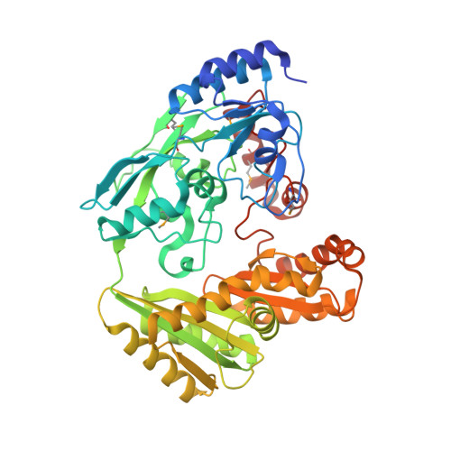 | |
UniProt | |||||
Entity Groups | |||||
| Sequence Clusters | 30% Identity50% Identity70% Identity90% Identity95% Identity100% Identity | ||||
| UniProt Group | Q6NAV4 | ||||
Sequence AnnotationsExpand | |||||
Reference Sequence | |||||
| Ligands 3 Unique | |||||
|---|---|---|---|---|---|
| ID | Chains | Name / Formula / InChI Key | 2D Diagram | 3D Interactions | |
| FAD Download:Ideal Coordinates CCD File | AA [auth E] FA [auth F] G [auth A] L [auth B] Q [auth C] | FLAVIN-ADENINE DINUCLEOTIDE C27 H33 N9 O15 P2 VWWQXMAJTJZDQX-UYBVJOGSSA-N |  | ||
| PO4 Download:Ideal Coordinates CCD File | CA [auth E] DA [auth E] EA [auth E] HA [auth F] I [auth A] | PHOSPHATE ION O4 P NBIIXXVUZAFLBC-UHFFFAOYSA-K |  | ||
| UNL Download:Ideal Coordinates CCD File | BA [auth E] GA [auth F] H [auth A] M [auth B] R [auth C] | Unknown ligand NBIIXXVUZAFLBC-UHFFFAOYSA-K | |||
| Modified Residues 1 Unique | |||||
|---|---|---|---|---|---|
| ID | Chains | Type | Formula | 2D Diagram | Parent |
| MSE Query on MSE | A, B, C, D, E A, B, C, D, E, F | L-PEPTIDE LINKING | C5 H11 N O2 Se |  | MET |
| Length ( Å ) | Angle ( ˚ ) |
|---|---|
| a = 146.085 | α = 90 |
| b = 250.728 | β = 90 |
| c = 251.711 | γ = 90 |
| Software Name | Purpose |
|---|---|
| SHELX | phasing |
| REFMAC | refinement |
| XSCALE | data scaling |
| PDB_EXTRACT | data extraction |
| XDS | data reduction |
| SHELXD | phasing |
| autoSHARP | phasing |


