3PJ7 | pdb_00003pj7

Crystal structure of far-red fluorescent protein Katushka crystallized at pH 8.5


Experimental Data Snapshot

  • Method: X-RAY DIFFRACTION
  • Resolution: 1.85 Å
  • R-Value Free: 
    0.252 (Depositor), 0.251 (DCC) 
  • R-Value Work: 
    0.196 (Depositor), 0.200 (DCC) 
  • R-Value Observed: 
    0.197 (Depositor) 

wwPDB Validation 3D Report Full Report

Validation slider image for 3PJ7

This is version 2.0 of the entry. See complete history

Literature

Crystallographic study of red fluorescent protein eqFP578 and its far-red variant Katushka reveals opposite pH-induced isomerization of chromophore.

Pletneva, N.V.Pletnev, V.Z.Shemiakina, I.I.Chudakov, D.M.Artemyev, I.Wlodawer, A.Dauter, Z.Pletnev, S.

(2011) Protein Sci 20: 1265-1274

  • DOI: https://doi.org/10.1002/pro.654
  • Primary Citation Related Structures: 
    3PIB, 3PJ5, 3PJ7, 3PJB

  • PubMed Abstract: 

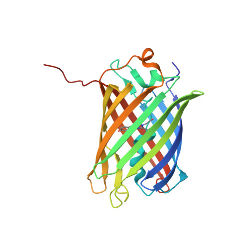
    The wild type red fluorescent protein eqFP578 (from sea anemone Entacmaea quadricolor, λ(ex) = 552 nm, λ(em) = 578 nm) and its bright far-red fluorescent variant Katushka (λ(ex) = 588 nm, λ(em) = 635 nm) are characterized by the pronounced pH dependence of their fluorescence. The crystal structures of eqFP578f (eqFP578 with two point mutations improving the protein folding) and Katushka have been determined at the resolution ranging from 1.15 to 1.85 Å at two pH values, corresponding to low and high level of fluorescence. The observed extinguishing of fluorescence upon reducing pH in eqFP578f and Katushka has been shown to be accompanied by the opposite trans-cis and cis-trans chromophore isomerization, respectively. Asn143, Ser158, His197 and Ser143, Leu174, and Arg197 have been shown to stabilize the respective trans and cis fluorescent states of the chromophores in eqFP578f and Katushka at higher pH. The cis state has been suggested as being primarily responsible for the observed far-red shift of the emission maximum of Katushka relative to that of eqFP578f.


  • Organizational Affiliation
    • Shemyakin-Ovchinnikov Institute of Bioorganic Chemistry, Russian Academy of Science, Moscow 117997, Miklukho-Maklaya 16/10, Russia. pletnevs@mail.nih.gov

Macromolecule Content 

  • Total Structure Weight: 103.96 kDa 
  • Atom Count: 7,633 
  • Modeled Residue Count: 892 
  • Deposited Residue Count: 916 
  • Unique protein chains: 1

Macromolecules

Find similar proteins by:|  3D Structure
Entity ID: 1
MoleculeChains  Sequence LengthOrganismDetailsImage
Red fluorescent protein eqFP578
A, B, C, D
229Entacmaea quadricolorMutation(s): 0 
UniProt
Find proteins for H3JQU6 (Entacmaea quadricolor)
Explore H3JQU6 
Go to UniProtKB:  H3JQU6
Entity Groups
Sequence Clusters30% Identity50% Identity70% Identity90% Identity95% Identity100% Identity
UniProt GroupH3JQU6
Sequence Annotations
Expand
Reference Sequence

Small Molecules

Modified Residues  1 Unique
IDChains TypeFormula2D DiagramParent
NRQ
Query on NRQ
A, B, C, D
L-PEPTIDE LINKINGC16 H17 N3 O4 SMET, TYR, GLY

Experimental Data & Validation

Experimental Data

  • Method: X-RAY DIFFRACTION
  • Resolution: 1.85 Å
  • R-Value Free:  0.252 (Depositor), 0.251 (DCC) 
  • R-Value Work:  0.196 (Depositor), 0.200 (DCC) 
  • R-Value Observed: 0.197 (Depositor) 
Space Group: I 41
Unit Cell:
Length ( Å )Angle ( ˚ )
a = 161.353α = 90
b = 161.353β = 90
c = 74.486γ = 90
Software Package:
Software NamePurpose
SCALEPACKdata scaling
REFMACrefinement
PDB_EXTRACTdata extraction
SERGUIdata collection
HKL-2000data reduction
HKL-2000data scaling
MOLREPphasing

Structure Validation

View Full Validation Report


Entry History 

Deposition Data

Revision History  (Full details and data files)

  • Version 1.0: 2011-05-25
    Type: Initial release
  • Version 1.1: 2011-07-13
    Changes: Version format compliance
  • Version 1.2: 2012-02-22
    Changes: Other
  • Version 1.3: 2025-03-26
    Changes: Advisory, Data collection, Database references, Derived calculations, Structure summary
  • Version 2.0: 2026-03-18
    Changes: Polymer sequence