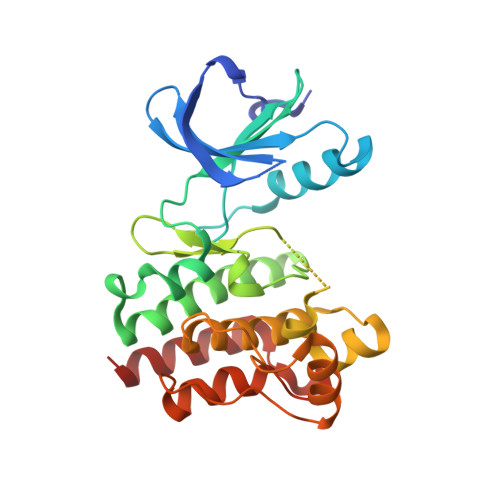
3PJ2

Crystal structure of BTK kinase domain complexed with 2-[4-(2-Diethylamino-ethoxy)-phenylamino]-6-(4-fluoro-phenoxy)-8-methyl-8H-pyrido[2,3-d]pyrimidin-7-one


Experimental Data Snapshot

  • Method: X-RAY DIFFRACTION
  • Resolution: 1.75 Å
  • R-Value Free: 0.263 
  • R-Value Work: 0.229 
  • R-Value Observed: 0.231 

Starting Model: experimental
View more details

wwPDB Validation   3D Report Full Report


Ligand Structure Quality Assessment 


This is version 1.2 of the entry. See complete history


Literature

Insights into the conformational flexibility of Bruton's tyrosine kinase from multiple ligand complex structures.

Kuglstatter, A.Wong, A.Tsing, S.Lee, S.W.Lou, Y.Villasenor, A.G.Bradshaw, J.M.Shaw, D.Barnett, J.W.Browner, M.F.

(2011) Protein Sci 20: 428-436

  • DOI: https://doi.org/10.1002/pro.575
  • Primary Citation of Related Structures:  
    3PIX, 3PIY, 3PIZ, 3PJ1, 3PJ2, 3PJ3

  • PubMed Abstract: 

    Bruton's tyrosine kinase (BTK) plays a key role in B cell receptor signaling and is considered a promising drug target for lymphoma and inflammatory diseases. We have determined the X-ray crystal structures of BTK kinase domain in complex with six inhibitors from distinct chemical classes. Five different BTK protein conformations are stabilized by the bound inhibitors, providing insights into the structural flexibility of the Gly-rich loop, helix C, the DFG sequence, and activation loop. The conformational changes occur independent of activation loop phosphorylation and do not correlate with the structurally unchanged WEI motif in the Src homology 2-kinase domain linker. Two novel activation loop conformations and an atypical DFG conformation are observed representing unique inactive states of BTK. Two regions within the activation loop are shown to structurally transform between 3(10)- and α-helices, one of which collapses into the adenosine-5'-triphosphate binding pocket. The first crystal structure of a Tec kinase family member in the pharmacologically important DFG-out conformation and bound to a type II kinase inhibitor is described. The different protein conformations observed provide insights into the structural flexibility of BTK, the molecular basis of its regulation, and the structure-based design of specific inhibitors.


  • Organizational Affiliation

    Roche Palo Alto, 3431 Hillview Avenue, Palo Alto, California 94304, USA. andreas.kuglstatter@roche.com


Macromolecules
Find similar proteins by:  (by identity cutoff)  |  3D Structure
Entity ID: 1
MoleculeChains Sequence LengthOrganismDetailsImage
Tyrosine-protein kinase BTK274Homo sapiensMutation(s): 0 
Gene Names: AGMX1ATKBPKBTK
EC: 2.7.10.2
UniProt & NIH Common Fund Data Resources
Find proteins for Q06187 (Homo sapiens)
Explore Q06187 
Go to UniProtKB:  Q06187
PHAROS:  Q06187
GTEx:  ENSG00000010671 
Entity Groups  
Sequence Clusters30% Identity50% Identity70% Identity90% Identity95% Identity100% Identity
UniProt GroupQ06187
Sequence Annotations
Expand
  • Reference Sequence
Small Molecules
Ligands 1 Unique
IDChains Name / Formula / InChI Key2D Diagram3D Interactions
04K
Query on 04K

Download Ideal Coordinates CCD File 
B [auth A]2-({4-[2-(diethylamino)ethoxy]phenyl}amino)-6-(4-fluorophenoxy)-8-methylpyrido[2,3-d]pyrimidin-7(8H)-one
C26 H28 F N5 O3
PAJOHXKWGHBVDS-UHFFFAOYSA-N
Binding Affinity Annotations 
IDSourceBinding Affinity
04K PDBBind:  3PJ2 IC50: 7200 (nM) from 1 assay(s)
Experimental Data & Validation

Experimental Data

  • Method: X-RAY DIFFRACTION
  • Resolution: 1.75 Å
  • R-Value Free: 0.263 
  • R-Value Work: 0.229 
  • R-Value Observed: 0.231 
  • Space Group: P 21 21 2
Unit Cell:
Length ( Å )Angle ( ˚ )
a = 71.356α = 90
b = 104.803β = 90
c = 38.002γ = 90
Software Package:
Software NamePurpose
ADSCdata collection
REFMACrefinement
DENZOdata reduction
SCALEPACKdata scaling
REFMACphasing

Structure Validation

View Full Validation Report


Ligand Structure Quality Assessment 


Entry History 

Deposition Data

Revision History  (Full details and data files)

  • Version 1.0: 2011-01-12
    Type: Initial release
  • Version 1.1: 2011-07-13
    Changes: Version format compliance
  • Version 1.2: 2023-09-06
    Changes: Data collection, Database references, Derived calculations, Refinement description