3PHB | pdb_00003phb

Crystal Structure of human purine nucleoside phosphorylase in complex with DADMe-ImmG


Experimental Data Snapshot

  • Method: X-RAY DIFFRACTION
  • Resolution: 2.30 Å
  • R-Value Free: 
    0.295 (Depositor), 0.295 (DCC) 
  • R-Value Work: 
    0.230 (Depositor), 0.230 (DCC) 
  • R-Value Observed: 
    0.233 (Depositor) 

wwPDB Validation 3D Report Full Report

Validation slider image for 3PHB

Ligand Structure Quality Assessment 


This is version 1.3 of the entry. See complete history

Literature

Crystal Structure of human purine nucleoside phosphorylase in complex with DADMe-ImmG

Ho, M.Cassera, M.B.Murkin, A.S.Almo, S.C.Schramm, V.L.

To be published.

Macromolecule Content 

  • Total Structure Weight: 219.7 kDa 
  • Atom Count: 13,732 
  • Modeled Residue Count: 1,718 
  • Deposited Residue Count: 1,944 
  • Unique protein chains: 1

Macromolecules

Find similar proteins by:|  3D Structure
Entity ID: 1
MoleculeChains  Sequence LengthOrganismDetailsImage
Purine nucleoside phosphorylase324Homo sapiensMutation(s): 0 
Gene Names: PNPNP
EC: 2.4.2.1
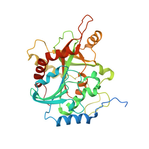
UniProt & NIH Common Fund Data Resources
Find proteins for P00491 (Homo sapiens)
Explore P00491 
Go to UniProtKB:  P00491
PHAROS:  P00491
GTEx:  ENSG00000198805 
Entity Groups
Sequence Clusters30% Identity50% Identity70% Identity90% Identity95% Identity100% Identity
UniProt GroupP00491
Sequence Annotations
Expand
Reference Sequence

Small Molecules

Ligands 2 Unique
IDChains Name / Formula / InChI Key2D Diagram3D Interactions
IM5

Query on IM5


Download:Ideal Coordinates CCD File
G [auth E]
J [auth Q]
N [auth S]
Q [auth T]
S [auth U]
G [auth E],
J [auth Q],
N [auth S],
Q [auth T],
S [auth U],
W [auth Y]
2-amino-7-{[(3R,4R)-3-hydroxy-4-(hydroxymethyl)pyrrolidin-1-yl]methyl}-3,5-dihydro-4H-pyrrolo[3,2-d]pyrimidin-4-one
C12 H17 N5 O3
GSPTUGDLYPMLCQ-SFYZADRCSA-N
PO4

Query on PO4


Download:Ideal Coordinates CCD File
H [auth E]
I [auth E]
K [auth Q]
L [auth Q]
M [auth S]
H [auth E],
I [auth E],
K [auth Q],
L [auth Q],
M [auth S],
O [auth S],
P [auth T],
R [auth T],
T [auth U],
U [auth Y],
V [auth Y],
X [auth Y]
PHOSPHATE ION
O4 P
NBIIXXVUZAFLBC-UHFFFAOYSA-K
Binding Affinity Annotations 
IDSourceBinding Affinity
IM5 BindingDB:  3PHB Ki: min: 7.00e-3, max: 7 (nM) from 3 assay(s)

Experimental Data & Validation

Experimental Data

  • Method: X-RAY DIFFRACTION
  • Resolution: 2.30 Å
  • R-Value Free:  0.295 (Depositor), 0.295 (DCC) 
  • R-Value Work:  0.230 (Depositor), 0.230 (DCC) 
  • R-Value Observed: 0.233 (Depositor) 
Space Group: C 1 2 1
Unit Cell:
Length ( Å )Angle ( ˚ )
a = 269.27α = 90
b = 52.632β = 90.33
c = 128.119γ = 90
Software Package:
Software NamePurpose
DENZOdata reduction
SCALEPACKdata scaling
REFMACrefinement
PDB_EXTRACTdata extraction

Structure Validation

View Full Validation Report


Ligand Structure Quality Assessment 


Entry History 

Deposition Data

Revision History  (Full details and data files)

  • Version 1.0: 2011-11-09
    Type: Initial release
  • Version 1.1: 2011-12-14
    Changes: Non-polymer description
  • Version 1.2: 2017-11-08
    Changes: Refinement description
  • Version 1.3: 2024-02-21
    Changes: Data collection, Database references, Derived calculations