3PGE | pdb_00003pge

Structure of sumoylated PCNA


Experimental Data Snapshot

  • Method: X-RAY DIFFRACTION
  • Resolution: 2.80 Å
  • R-Value Free: 
    0.253 (Depositor), 0.296 (DCC) 
  • R-Value Work: 
    0.231 (Depositor), 0.279 (DCC) 
  • R-Value Observed: 
    0.232 (Depositor) 

Starting Model: experimental
View more details

wwPDB Validation 3D Report Full Report

Validation slider image for 3PGE

This is version 1.3 of the entry. See complete history

Literature

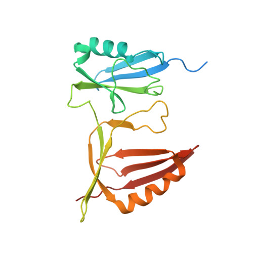
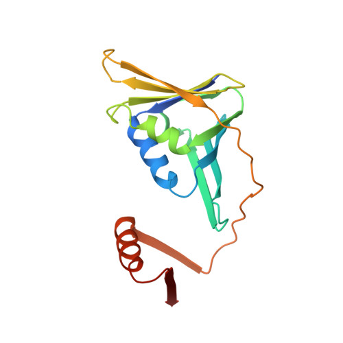
Crystal Structure of SUMO-Modified Proliferating Cell Nuclear Antigen.

Freudenthal, B.D.Brogie, J.E.Gakhar, L.Kondratick, C.M.Washington, M.T.

(2011) J Mol Biology 406: 9-17

  • DOI: https://doi.org/10.1016/j.jmb.2010.12.015
  • Primary Citation Related Structures: 
    3PGE

  • PubMed Abstract: 

    Eukaryotic proliferating cell nuclear antigen (PCNA) is a replication accessory protein that functions in DNA replication, repair, and recombination. The various functions of PCNA are regulated by posttranslational modifications including mono-ubiquitylation, which promotes translesion synthesis, and sumoylation, which inhibits recombination. To understand how SUMO modification regulates PCNA, we generated a split SUMO-modified PCNA protein and showed that it supports cell viability and stimulates DNA polymerase δ activity. We then determined its X-ray crystal structure and found that SUMO occupies a position on the back face of the PCNA ring, which is distinct from the position occupied by ubiquitin in the structure of ubiquitin-modified PCNA. We propose that the back of PCNA has evolved to be a site of regulation that can be easily modified without disrupting ongoing reactions on the front of PCNA, such as normal DNA replication. Moreover, these modifications likely allow PCNA to function as a tool belt, whereby proteins can be recruited to the replication machinery via the back of PCNA and be held in reserve until needed.


  • Organizational Affiliation
    • Department of Biochemistry, University of Iowa College of Medicine, Iowa City, IA 52242-1109, USA.

Macromolecule Content 

  • Total Structure Weight: 42.03 kDa 
  • Atom Count: 2,647 
  • Modeled Residue Count: 335 
  • Deposited Residue Count: 371 
  • Unique protein chains: 2

Macromolecules

Find similar proteins by:|  3D Structure
Entity ID: 1
MoleculeChains  Sequence LengthOrganismDetailsImage
SUMO-modified proliferating cell nuclear antigen200Saccharomyces cerevisiaeMutation(s): 0 
UniProt
Find proteins for P15873 (Saccharomyces cerevisiae (strain ATCC 204508 / S288c))
Explore P15873 
Go to UniProtKB:  P15873
Find proteins for Q12306 (Saccharomyces cerevisiae (strain ATCC 204508 / S288c))
Explore Q12306 
Go to UniProtKB:  Q12306
Entity Groups
Sequence Clusters30% Identity50% Identity70% Identity90% Identity95% Identity100% Identity
UniProt GroupsQ12306P15873
Sequence Annotations
Expand
Reference Sequence
Find similar proteins by:|  3D Structure
Entity ID: 2
MoleculeChains  Sequence LengthOrganismDetailsImage
Proliferating cell nuclear antigen171Saccharomyces cerevisiaeMutation(s): 0 
UniProt
Find proteins for P15873 (Saccharomyces cerevisiae (strain ATCC 204508 / S288c))
Explore P15873 
Go to UniProtKB:  P15873
Entity Groups
Sequence Clusters30% Identity50% Identity70% Identity90% Identity95% Identity100% Identity
UniProt GroupP15873
Sequence Annotations
Expand
Reference Sequence

Experimental Data & Validation

Experimental Data

  • Method: X-RAY DIFFRACTION
  • Resolution: 2.80 Å
  • R-Value Free:  0.253 (Depositor), 0.296 (DCC) 
  • R-Value Work:  0.231 (Depositor), 0.279 (DCC) 
  • R-Value Observed: 0.232 (Depositor) 
Space Group: F 4 3 2
Unit Cell:
Length ( Å )Angle ( ˚ )
a = 268.816α = 90
b = 268.816β = 90
c = 268.816γ = 90
Software Package:
Software NamePurpose
StructureStudiodata collection
PHASERphasing
PHENIXrefinement
d*TREKdata reduction
d*TREKdata scaling

Structure Validation

View Full Validation Report


Entry History 

Deposition Data

Revision History  (Full details and data files)

  • Version 1.0: 2010-12-29
    Type: Initial release
  • Version 1.1: 2011-07-13
    Changes: Version format compliance
  • Version 1.2: 2017-08-02
    Changes: Refinement description, Source and taxonomy
  • Version 1.3: 2023-09-06
    Changes: Data collection, Database references, Refinement description